

Using sea urchin gametes and zygotes to investigate centrosome duplication
- PMID: 27602205
- PMCID: PMC5011938
- DOI: 10.1186/s13630-016-0043-3
Using sea urchin gametes and zygotes to investigate centrosome duplication
Abstract
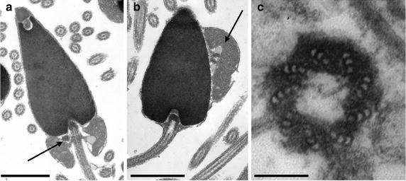
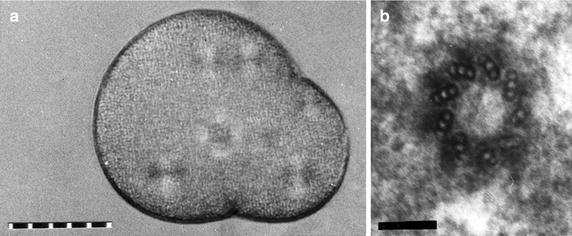
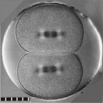
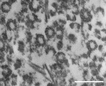
Centriole structure and function in the sea urchin zygote parallel those in mammalian somatic cells. Here, I briefly introduce the properties and attributes of the sea urchin system that make it an attractive platform for the study of centrosome and centriole duplication. These attributes apply to all echinoderms readily available from commercial suppliers: sea urchins, sand dollars, and starfish. I list some of the practical aspects of the system that make it a cost- and time-effective system for experimental work and then list properties that are a "tool kit" that can be used to conduct studies that would not be practical, or in some cases not possible, with mammalian somatic cells. Since centrioles organize and localize the pericentriolar material that nucleates the astral arrays of microtubules (Bobinnec et al. in J Cell Biol 143(6):1575-1589, 1998), the pattern of aster duplication over several cell cycles can be used as a reliable measure for centriole duplication (Sluder and Rieder in J Cell Biol 100(3):887-896, 1985). Descriptions of the methods my laboratory has used to handle and image echinoderm zygotes are reviewed in Sluder et al. (Methods Cell Biol 61:439-472, 1999). Also included is a bibliography of papers that describe additional methods.
Keywords: Centriole; Centrosome; Duplication; Echinoderm; Egg; Sea urchin; Sperm; Zygote.
Figures
Similar articles
-
Reproductive maternal centrosomes are cast off into polar bodies during maturation division in starfish oocytes.Exp Cell Res. 2001 Sep 10;269(1):130-9. doi: 10.1006/excr.2001.5305. Exp Cell Res. 2001. PMID: 11525646
-
Mitosis in the human embryo: the vital role of the sperm centrosome (centriole).Histol Histopathol. 1997 Jul;12(3):827-56. Histol Histopathol. 1997. PMID: 9225167 Review.
-
Characterization of human gamete centrosomes for assisted reproduction.Ital J Anat Embryol. 2001;106(2 Suppl 2):61-73. Ital J Anat Embryol. 2001. PMID: 11732597 Review.
-
Cyclin E in centrosome duplication and reduplication in sea urchin zygotes.J Cell Physiol. 2008 Dec;217(3):626-31. doi: 10.1002/jcp.21531. J Cell Physiol. 2008. PMID: 18651565 Free PMC article.
-
Reproductive capacity of sea urchin centrosomes without centrioles.Cell Motil Cytoskeleton. 1989;13(4):264-73. doi: 10.1002/cm.970130405. Cell Motil Cytoskeleton. 1989. PMID: 2776224
Cited by
-
The Evolution of Centriole Structure: Heterochrony, Neoteny, and Hypermorphosis.Results Probl Cell Differ. 2019;67:3-15. doi: 10.1007/978-3-030-23173-6_1. Results Probl Cell Differ. 2019. PMID: 31435789 Free PMC article. Review.
-
It takes two (centrioles) to tango.Reproduction. 2019 Feb;157(2):R33-R51. doi: 10.1530/REP-18-0350. Reproduction. 2019. PMID: 30496124 Free PMC article. Review.
-
Sophisticated lessons from simple organisms: appreciating the value of curiosity-driven research.Dis Model Mech. 2017 Dec 19;10(12):1381-1389. doi: 10.1242/dmm.031203. Dis Model Mech. 2017. PMID: 29259023 Free PMC article. Review.
-
The impact of propranolol, 17α-ethinylestradiol, and gemfibrozil on early life stages of marine organisms: effects and risk assessment.Environ Sci Pollut Res Int. 2018 Nov;25(32):32196-32209. doi: 10.1007/s11356-018-3185-6. Epub 2018 Sep 16. Environ Sci Pollut Res Int. 2018. PMID: 30220067
-
New techniques for creating parthenogenetic larvae of the sea urchin Lytechinus pictus for gene expression studies.Dev Dyn. 2021 Dec;250(12):1828-1833. doi: 10.1002/dvdy.377. Epub 2021 Jun 22. Dev Dyn. 2021. PMID: 34042247 Free PMC article.
References
Publication types
Grants and funding
LinkOut - more resources
Full Text Sources
Other Literature Sources
Miscellaneous
