

Expression of microphthalmia transcription factor, S100 protein, and HMB-45 in malignant melanoma and pigmented nevi
- PMID: 27602212
- PMCID: PMC4998223
- DOI: 10.3892/br.2016.732
Expression of microphthalmia transcription factor, S100 protein, and HMB-45 in malignant melanoma and pigmented nevi
Abstract
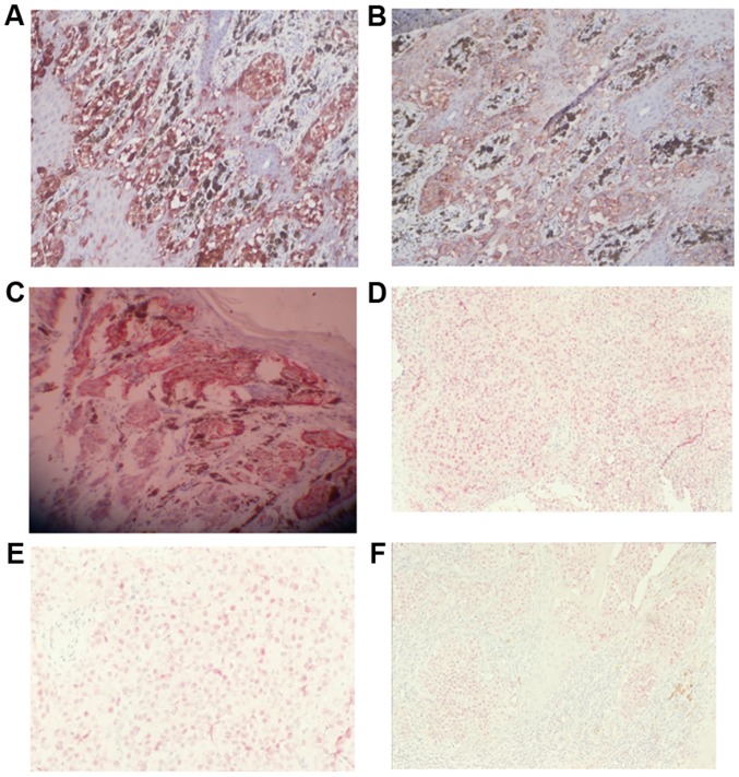
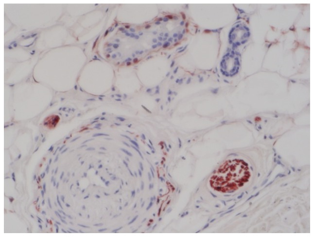
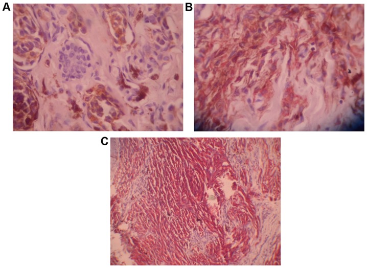
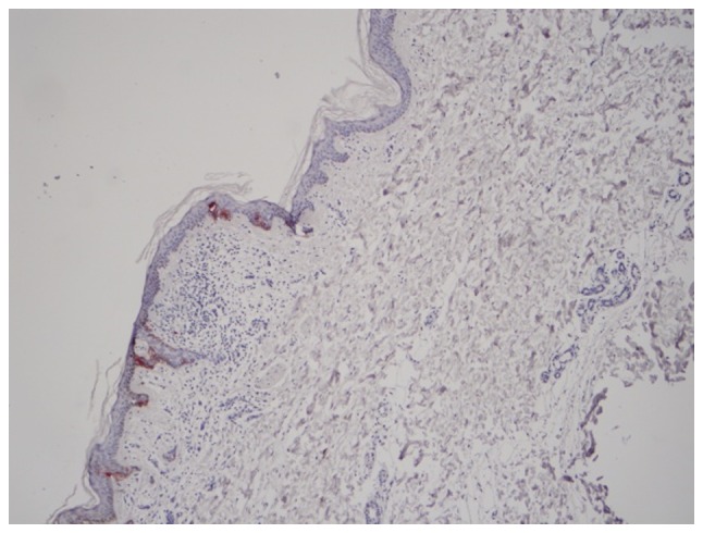
Malignant melanoma (MM) is a type of malignant tumor, which originates from neural crest melanocytes. MM progresses rapidly and results in a high mortality rate. The present study aims to investigate the expression of microphthalmia transcription factor (MITF), the S100 protein, and HMB-45 in MM and pigmented nevi. A total of 32 MM samples (including three skin metastasis, three lymph node metastasis and two spindle cell MM samples), two Spitz nevus samples, four pigmented nevus samples and two blue nevus samples were collected. The expression levels of S100 protein, HMB-45, and MITF were observed via immunostaining. The S100 protein exhibited high positive rates in MM and pigment disorders (96.7 and 100%, respectively), but with low specificity. The S100 protein was also expressed in fibroblasts, myoepithelial cells, histocytes and Langerhans cells in normal skin samples. HMB-45 had high specificity. Its positive expression was only confined to MM cells and junctional nevus cells. Furthermore, HMB-45 was not expressed in melanocytes in the normal tissue samples around the tumor or in the benign intradermal nevus cells. MITF exhibited high specificity and high sensitivity. It was expressed in the nuclei of melanocytes, MM cells and nevus cells. It was observed to be strongly expressed in metastatic MM and spindle cell MMs. Thus, MITF may present as a specific immunomarker for the diagnosis and differential diagnosis of MM.
Keywords: Spitz nevus; lymph node metastasis; malignant melanoma.
Figures
Similar articles
-
Microphthalmia transcription factor expression in cutaneous benign, malignant melanocytic, and nonmelanocytic tumors.Am J Surg Pathol. 2001 Jan;25(1):51-7. doi: 10.1097/00000478-200101000-00005. Am J Surg Pathol. 2001. PMID: 11145251
-
HMB-45 staining in benign and malignant melanocytic lesions. A reflection of cellular activation.Am J Dermatopathol. 1991 Dec;13(6):543-50. doi: 10.1097/00000372-199113060-00004. Am J Dermatopathol. 1991. PMID: 1725245
-
Expression of Melan-A in Spitz, pigmented spindle cell nevi, and congenital nevi: comparative immunohistochemical study.Pediatr Dev Pathol. 2000 Jan-Feb;3(1):36-9. doi: 10.1007/pl00021520. Pediatr Dev Pathol. 2000. PMID: 10644168
-
Intraoral Spitz nevus: case report and literature review.Oral Surg Oral Med Oral Pathol Oral Radiol. 2014 Apr;117(4):e320-4. doi: 10.1016/j.oooo.2013.07.025. Epub 2013 Oct 16. Oral Surg Oral Med Oral Pathol Oral Radiol. 2014. PMID: 24139994 Review.
-
[Spitz nevus and Reed nevus: simulating melanoma in adults].Pathologe. 1998 Nov;19(6):403-11. doi: 10.1007/s002920050304. Pathologe. 1998. PMID: 9885003 Review. German.
Cited by
-
Clinical and immunohistochemical analysis of the verrucous and non-verrucous divided nevus of the eyelids.BMC Ophthalmol. 2022 Sep 3;22(1):358. doi: 10.1186/s12886-022-02582-w. BMC Ophthalmol. 2022. PMID: 36057574 Free PMC article.
-
The Importance of Immunohistochemistry in the Evaluation of Tumor Depth of Primary Cutaneous Melanoma.Diagnostics (Basel). 2023 Mar 7;13(6):1020. doi: 10.3390/diagnostics13061020. Diagnostics (Basel). 2023. PMID: 36980327 Free PMC article.
-
Primary thoracolumbar intraspinal malignant melanoma: A case report.World J Clin Cases. 2024 Jun 6;12(16):2904-2910. doi: 10.12998/wjcc.v12.i16.2904. World J Clin Cases. 2024. PMID: 38899297 Free PMC article.
-
Cutaneous Melanoma: A Review of Multifactorial Pathogenesis, Immunohistochemistry, and Emerging Biomarkers for Early Detection and Management.Int J Mol Sci. 2023 Nov 1;24(21):15881. doi: 10.3390/ijms242115881. Int J Mol Sci. 2023. PMID: 37958863 Free PMC article. Review.
-
Primary intramedullary melanoma of lumbar spinal cord: A case report.World J Clin Cases. 2021 Apr 6;9(10):2352-2356. doi: 10.12998/wjcc.v9.i10.2352. World J Clin Cases. 2021. PMID: 33869613 Free PMC article.
References
-
- Rastrelli M, Tropea S, Rossi CR, Alaibac M. Melanoma: Epidemiology, risk factors, pathogenesis, diagnosis and classification. Vivo. 2014;28:1005–1011. - PubMed
-
- National Collaborating Centre for Cancer. Melanoma: Assessment and management. National Institute for Health and Care Excellence; London: 2015. - PubMed
-
- Scolyer RA, Li LX, McCarthy SW, Shaw HM, Stretch JR, Sharma R, Thompson JF. Immunohistochemical stains fail to increase the detection rate of micrometastatic melanoma in completion regional lymph node dissection specimens. Melanoma Res. 2004;14:263–268. doi: 10.1097/01.cmr.0000136708.90534.71. - DOI - PubMed
LinkOut - more resources
Full Text Sources
Other Literature Sources