

Myxoid chondrosarcoma of the mandible in a 22-year-old man: A case report
- PMID: 27602220
- PMCID: PMC4998218
- DOI: 10.3892/mco.2016.939
Myxoid chondrosarcoma of the mandible in a 22-year-old man: A case report
Abstract
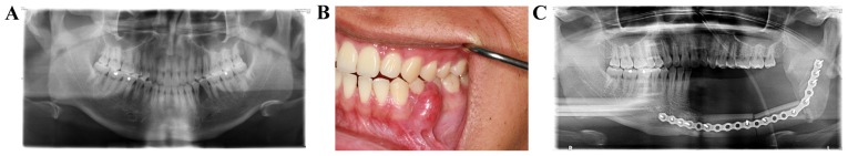
This study describes a case of myxoid chondrosarcoma of the mandible in a 22-year-old male patient. A tumour in the buccal gingiva of the lower left premolar region had been identified 2 years earlier. Whole-jaw panoramic radiographs showed a hypodense shadow in the mesiodistal area near the roots of teeth 34 and 35. A maxillofacial computed tomography scan revealed a mass in the lower left premolar soft tissue, with a shadow indicating bone destruction, a clear boundary and uniform density. The preliminary diagnosis at the outpatient department was 34-35 epulis. The patient underwent surgery for 34-35 gingival tumour resection, 34 and 35 extraction, and 34 and 35 immediate implantation. The postoperative pathological examination revealed a cellular type extraskeletal myxoid chondrosarcoma of the lower left mandible. Under general anaesthesia, the patient underwent lower left mandibular block and segmental resection, submandibular triangle dissection and vessel disassociation, and musculocutaneous flap repair in the oral and maxillofacial defect area. After 9 months of follow-up, the patient had no complaints of discomfort, and tumour recurrence was not observed on imaging examinations.
Keywords: extraskeletal myxoid chondrosarcoma; mandible; soft tissue.
Figures

Similar articles
-
Extraskeletal myxoid chondrosarcoma of the gingival: a rare case report and review of the literature.Diagn Pathol. 2023 Sep 13;18(1):103. doi: 10.1186/s13000-023-01390-0. Diagn Pathol. 2023. PMID: 37705036 Free PMC article. Review.
-
Diagnosis of extraskeletal myxoid chondrosarcoma in the thigh using EWSR1-NR4A3 gene fusion: a case report.J Med Case Rep. 2016 Nov 10;10(1):321. doi: 10.1186/s13256-016-1113-2. J Med Case Rep. 2016. PMID: 27832806 Free PMC article.
-
Breast Metastasis of Extraskeletal Myxoid Chondrosarcoma: A Case Report.Am J Case Rep. 2015 Jun 30;16:406-14. doi: 10.12659/AJCR.894804. Am J Case Rep. 2015. PMID: 26125202 Free PMC article.
-
Extraskeletal myxoid chondrosarcoma of the perineum.Orthopedics. 2009 Mar;32(3):216. Orthopedics. 2009. PMID: 19309044
-
Myxofibrosarcoma of the mandible: a case report and review of the literature.BMC Oral Health. 2020 Apr 16;20(1):113. doi: 10.1186/s12903-020-01094-7. BMC Oral Health. 2020. PMID: 32299394 Free PMC article. Review.
Cited by
-
Juxtacortical Mandibular Chondrosarcoma during pregnancy: A case report.J Clin Exp Dent. 2017 May 1;9(5):e723-e725. doi: 10.4317/jced.53630. eCollection 2017 May. J Clin Exp Dent. 2017. PMID: 28512553 Free PMC article.
-
Extraskeletal Myxoid Chondrosarcoma of Floor of Mouth-A Rare Case Report and Review of Literature.Indian J Otolaryngol Head Neck Surg. 2024 Feb;76(1):1290-1297. doi: 10.1007/s12070-023-04271-6. Epub 2023 Oct 14. Indian J Otolaryngol Head Neck Surg. 2024. PMID: 38440485 Free PMC article.
-
Extraskeletal Chondrosarcoma: Long-term Follow-up of a Patient with Metastatic Disease.Cureus. 2018 May 30;10(5):e2709. doi: 10.7759/cureus.2709. Cureus. 2018. PMID: 30065902 Free PMC article.
-
Extraskeletal myxoid chondrosarcoma of the gingival: a rare case report and review of the literature.Diagn Pathol. 2023 Sep 13;18(1):103. doi: 10.1186/s13000-023-01390-0. Diagn Pathol. 2023. PMID: 37705036 Free PMC article. Review.
References
-
- Fletcher CDM, Bridge JA, Hogendoorn P, Mertens F, editors. WHO Classification of Tumours. 4th. Vol. 5. IARC press; Lyon: 2013. WHO classification of tumours of soft tissue and bone; pp. 223–224.
LinkOut - more resources
Full Text Sources
Other Literature Sources