Piwil2 is reactivated by HPV oncoproteins and initiates cell reprogramming via epigenetic regulation during cervical cancer tumorigenesis
- PMID: 27602489
- PMCID: PMC5323100
- DOI: 10.18632/oncotarget.11810
Piwil2 is reactivated by HPV oncoproteins and initiates cell reprogramming via epigenetic regulation during cervical cancer tumorigenesis
Abstract
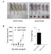
The human papillomavirus (HPV) oncoproteins E6 and E7 are risk factors that are primarily responsible for the initiation and progression of cervical cancer, and they play a key role in immortalization and transformation by reprogramming differentiating host epithelial cells. It is unclear how cervical epithelial cells transform into tumor-initiating cells (TICs). Here, we observed that the germ stem cell protein Piwil2 is expressed in pre-cancerous and malignant lesions of the cervix and cervical cancer cell lines with the exception of the non-HPV-infected C33a cell line. Knockdown of Piwil2 by shRNA led to a marked reduction in proliferation and colony formation, in vivo tumorigenicity, chemo-resistance, and the proportion of cancer stem-like cells. In contrast, Piwil2 overexpression induced malignant transformation of HaCaT cells and the acquisition of tumor-initiating capabilities. Gene-set enrichment analysis revealed embryonic stem cell (ESC) identity, malignant biological behavior, and specifically, activation targets of the cell reprogramming factors c-Myc, Klf4, Nanog, Oct4, and Sox2 in Piwil2-overexpressing HaCaT cells. We further confirmed that E6 and E7 reactivated Piwil2 and that E6 and E7 overexpression resulted in a similar gene-set enrichment pattern as Piwil2 overexpression in HaCaT cells. Moreover, Piwil2 overexpression or E6 and E7 activation induced H3K9 acetylation but reduced H3K9 trimethylation, which contributed to the epigenetic reprogramming and ESC signature maintenance, as predicted previously. Our study demonstrates that Piwil2, reactivated by the HPV oncoproteins E6 and E7, plays an essential role in the transformation of cervical epithelial cells to TICs via epigenetics-based cell reprogramming.
Keywords: HPV oncoprotein; Piwil2; cell reprogramming; cervical cancer; tumor initiating cell.
Conflict of interest statement
The authors declare no conflicts of interest.
Figures


Similar articles
-
HPV-16 E6/E7 promotes cell migration and invasion in cervical cancer via regulating cadherin switch in vitro and in vivo.Arch Gynecol Obstet. 2015 Dec;292(6):1345-54. doi: 10.1007/s00404-015-3787-x. Epub 2015 Jun 21. Arch Gynecol Obstet. 2015. PMID: 26093522
-
CBP-mediated Wnt3a/β-catenin signaling promotes cervical oncogenesis initiated by Piwil2.Neoplasia. 2021 Jan;23(1):1-11. doi: 10.1016/j.neo.2020.10.013. Epub 2020 Nov 13. Neoplasia. 2021. PMID: 33190089 Free PMC article.
-
HPV16 E6/E7-mediated regulation of PiwiL1 expression induces tumorigenesis in cervical cancer cells.Cell Oncol (Dordr). 2024 Jun;47(3):917-937. doi: 10.1007/s13402-023-00904-8. Epub 2023 Dec 1. Cell Oncol (Dordr). 2024. PMID: 38036929
-
Molecular mechanisms underlying human papillomavirus E6 and E7 oncoprotein-induced cell transformation.Mutat Res Rev Mutat Res. 2017 Apr-Jun;772:23-35. doi: 10.1016/j.mrrev.2016.08.001. Epub 2016 Aug 5. Mutat Res Rev Mutat Res. 2017. PMID: 28528687 Review.
-
[Molecular basis of cervical carcinogenesis by high-risk human papillomaviruses].Uirusu. 2008 Dec;58(2):141-54. doi: 10.2222/jsv.58.141. Uirusu. 2008. PMID: 19374192 Review. Japanese.
Cited by
-
Unveiling the atlas of associations between 1,400 plasma metabolites and 24 tumors: Mendelian randomization analyses.Transl Cancer Res. 2024 Sep 30;13(9):4938-4956. doi: 10.21037/tcr-24-359. Epub 2024 Sep 9. Transl Cancer Res. 2024. PMID: 39430859 Free PMC article.
-
Advances and challenges in the use of liquid biopsy in gynaecological oncology.Heliyon. 2024 Oct 15;10(20):e39148. doi: 10.1016/j.heliyon.2024.e39148. eCollection 2024 Oct 30. Heliyon. 2024. PMID: 39492906 Free PMC article. Review.
-
LINC00673 rs11655237 Polymorphism Is Associated With Increased Risk of Cervical Cancer in a Chinese Population.Cancer Control. 2018 Jan-Dec;25(1):1073274818803942. doi: 10.1177/1073274818803942. Cancer Control. 2018. PMID: 30286619 Free PMC article.
-
PIWIL-2 and piRNAs are regularly expressed in epithelia of the skin and their expression is related to differentiation.Arch Dermatol Res. 2020 Dec;312(10):705-714. doi: 10.1007/s00403-020-02052-7. Epub 2020 Mar 12. Arch Dermatol Res. 2020. PMID: 32166374 Free PMC article.
-
The necessity of ZSCAN4 for preimplantation development and gene expression of bovine embryos.J Reprod Dev. 2019 Aug 9;65(4):319-326. doi: 10.1262/jrd.2019-039. Epub 2019 Apr 25. J Reprod Dev. 2019. PMID: 31019155 Free PMC article.
References
-
- Song JJ, Smith SK, Hannon GJ, Joshua-Tor L. Crystal structure of Argonaute and its implications for RISC slicer activity. Science. 2004;305:1434–1437. - PubMed
-
- Homolka D, Pandey RR, Goriaux C, Brasset E, Vaury C, Sachidanandam R, Fauvarque MO, Pillai RS. PIWI Slicing and RNA Elements in Precursors Instruct Directional Primary piRNA Biogenesis. Cell Rep. 2015;12:418–428. - PubMed
MeSH terms
Substances
LinkOut - more resources
Full Text Sources
Other Literature Sources
Medical
Research Materials

