. 2016 Sep 6;111(5):1008-13.
doi: 10.1016/j.bpj.2016.07.043.

The Screw-Like Movement of a Gliding Bacterium Is Powered by Spiral Motion of Cell-Surface Adhesins

Affiliations

The Screw-Like Movement of a Gliding Bacterium Is Powered by Spiral Motion of Cell-Surface Adhesins

Abhishek Shrivastava et al. Biophys J. .

Abstract

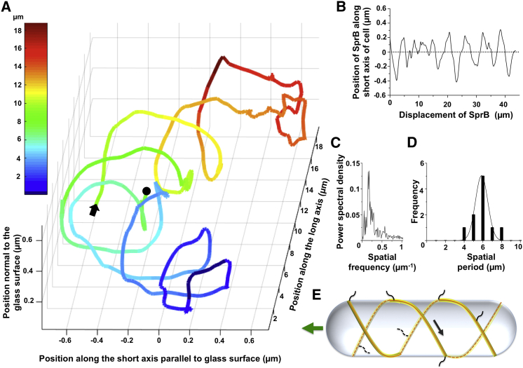
Flavobacterium johnsoniae, a rod-shaped bacterium, glides over surfaces at speeds of ∼2 μm/s. The propulsion of a cell-surface adhesin, SprB, is known to enable gliding. We used cephalexin to generate elongated cells with irregular shapes and followed their displacement in three dimensions. These cells rolled about their long axes as they moved forward, following a right-handed trajectory. We coated gold nanoparticles with an SprB antibody and tracked them in three dimensions in an evanescent field where the nanoparticles appeared brighter when they were closer to the glass. The nanoparticles followed a right-handed spiral trajectory on the surface of the cell. Thus, if SprB were to adhere to the glass rather than to a nanoparticle, the cell would move forward along a right-handed trajectory, as observed, but in a direction opposite to that of the nanoparticle.


Figures

Figure 1
Figure 1
Cells move sideways as they glide forward. (A) The gliding speed of 53 elongated cells was independent of cell length. Speed was plotted as dots and a linear regression was fit to the data. The coefficient of correlation was 0.03. (B) The distance of the pole near the bend of one cell, shown in Movie S1, from the long axis changed periodically with the distance traveled by the cell along its long axis. This distance was calculated using the method drawn in Fig. S2. (C) The spatial frequency of the displacement of the cell in Movie S1 moving along its long axis was 0.15 μm−1 and the spatial period was 6.6 μm. (D) Frequency distribution of the spatial periods for 31 cells. The average spatial period was 6.6 ± 1.2 μm.
Figure 2
Figure 2
Three-dimensional tracking reveals a screw-like mechanism. (A) Diagrammatic representation of a model to test whether a cell rolls like a screw or rocks back and forth. Conformations a, b, c, and d of a bent cell are depicted by black lines. Conformations b and d represent the image of the bent end of the cell when that segment lies in the microscope’s image plane (z = 0). The dashed line in (i) represents the image of the bent end of the cell when that segment is above the image plane (z > 0). The part of this segment that extends beyond the edge of the depth of field (represented by the white line) is not visible using the microscope with the high NA objective described in Materials and Methods. The dashed line in (ii) shows the part of the cell that is raised above the image plane by the cell rotation but remains within the depth of field. (B) Binary logic for the model. As a result of the decrease in the overall length of the cell image, a = 0, and b, c, and d (in which the imaged lengths are the same) = 1. Sequences for screw-like motion and rocking back and forth are boxed and the outputs for both scenarios are plotted. The periodicities for these plots are β = 6.6 and 3.3 μm, respectively. (C) Actual imaged cell length plotted as a function of distance traveled by a cell. This curve is similar to that obtained for the simulation of a cell rolling like a screw. (D) The frequency distribution of 32 β-values has a peak of 6.95 ± 1.11 μm, which is similar to the simulated frequency distribution of 32 β-values calculated from five cells that rolled like a screw while gliding forward.
Figure 3
Figure 3
SprB moves along a spiral trajectory. (A) A three-dimensional track of SprB appears as an irregular right-handed spiral. An arrow and a dot indicate the beginning and end of the track, respectively. The data shown are from cell 1 in Movie S3. Similar tracks were visualized for cells 2 and 3 from Movie S3 and are shown in Fig. S5. The color of the track represents the position of SprB along the long axis of the cell. (B) The position of one gold nanoparticle attached to SprB (cell 1, Movie S3) changed periodically along the short axis of the cell as the nanoparticle moved along the long axis of the cell. (C) The spatial frequency of movement of one gold nanoparticle (cell 1, Movie S3) along the short axis of one cell was 0.165 μm−1 and the spatial period was 6.06 μm. (D) Frequency distribution of the spatial periods for 10 cells. The average spatial period was 5.9 ± 0.7 μm. (E) A screw-like mechanism for gliding: a track (orange) remains fixed relative to the cell. SprB (black) and a tread (yellow) are in motion relative to the cell surface. The arrow shows the right-handed motion of the SprB and tread. Interaction of SprB with an external substratum results in forward motion and right-handed rotation of the cell body.

References

    1. Skerker J.M., Berg H.C. Direct observation of extension and retraction of type IV pili. Proc. Natl. Acad. Sci. USA. 2001;98:6901–6904. - PMC - PubMed
    1. Friedrich C., Bulyha I., Søgaard-Andersen L. Outside-in assembly pathway of the type IV pilus system in Myxococcus xanthus. J. Bacteriol. 2014;196:378–390. - PMC - PubMed
    1. Biais N., Higashi D.L., Sheetz M.P. Force-dependent polymorphism in type IV pili reveals hidden epitopes. Proc. Natl. Acad. Sci. USA. 2010;107:11358–11363. - PMC - PubMed
    1. Shrivastava A., Berg H.C. Towards a model for Flavobacterium gliding. Curr. Opin. Microbiol. 2015;28:93–97. - PMC - PubMed
    1. Islam S.T., Mignot T. The mysterious nature of bacterial surface (gliding) motility: a focal adhesion-based mechanism in Myxococcus xanthus. Semin. Cell Dev. Biol. 2015;46:143–154. - PubMed

LinkOut - more resources