

Epithelialization of mouse ovarian tumor cells originating in the fallopian tube stroma
- PMID: 27602775
- PMCID: PMC5323216
- DOI: 10.18632/oncotarget.11808
Epithelialization of mouse ovarian tumor cells originating in the fallopian tube stroma
Abstract
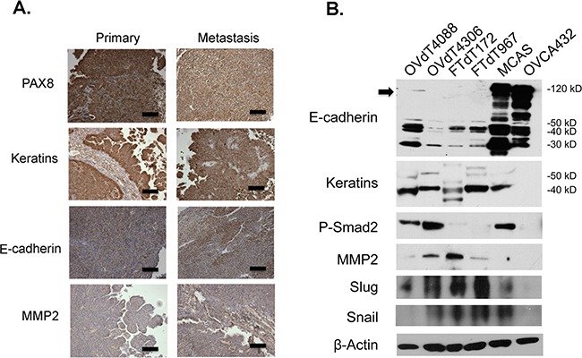
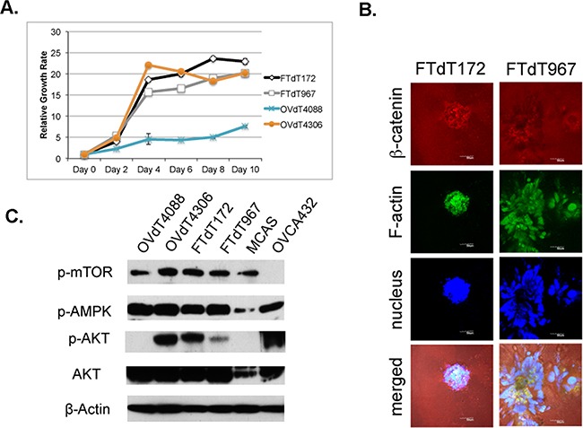
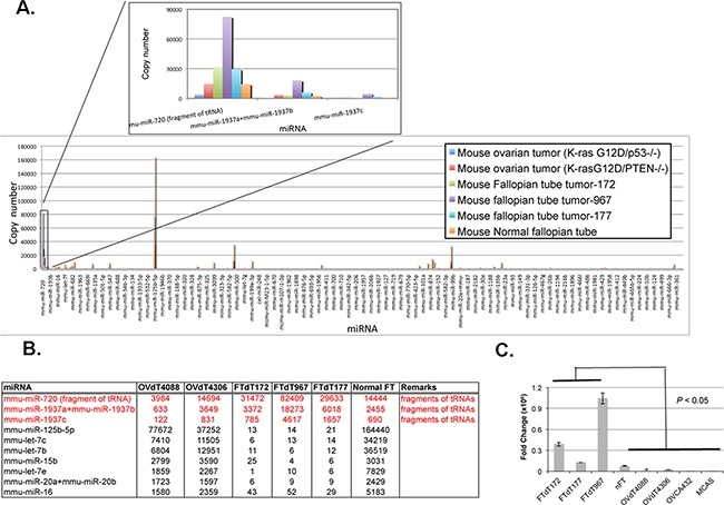
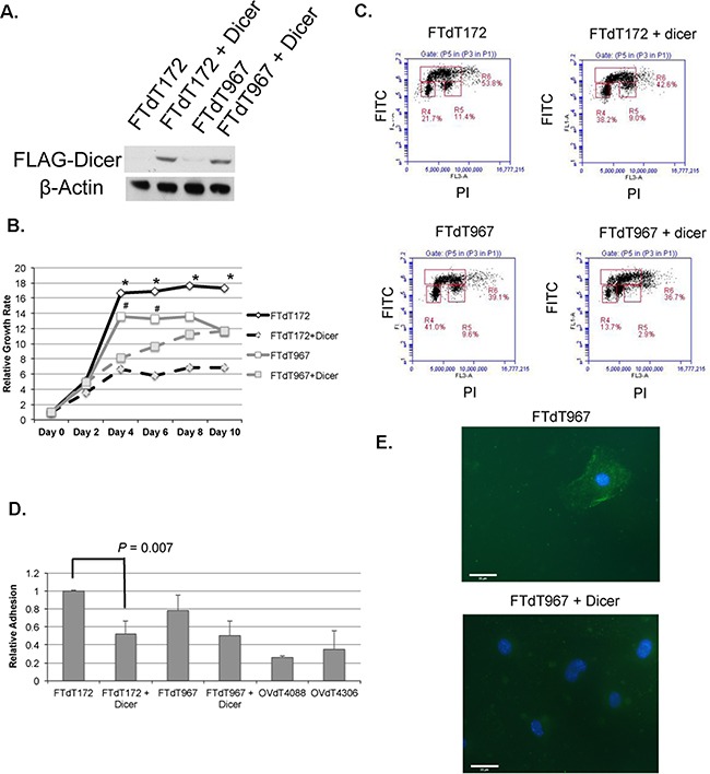
Epithelial ovarian carcinoma accounts for 90% of all ovarian cancer and is the most deadly gynecologic malignancy. Recent studies have suggested that fallopian tube fimbriae can be the origin of cells for high-grade serous subtype of epithelial ovarian carcinoma (HGSOC). A mouse HGSOC model with conditional Dicer-Pten double knockout (Dicer-Pten DKO) developed primary tumors, intriguingly, from the fallopian tube stroma. We examined the growth and epithelial phenotypes of the Dicer-Pten DKO mouse tumor cells contributable by each gene knockout. Unlike human ovarian epithelial cancer cells that expressed full-length E-cadherin, the Dicer-Pten DKO stromal tumor cells expressed cleaved E-cadherin fragments and metalloproteinase 2, a mixture of epithelial and mesenchymal markers. Although the Dicer-Pten DKO tumor cells lost the expression of mature microRNAs as expected, they showed high levels of tRNA fragment expression and enhanced AKT activation due to the loss of PTEN function. Introduction of a Dicer1-expressing construct into the DKO mouse tumor cells significantly reduced DNA synthesis and the cell growth rate, with concurrent diminished adhesion and ZO1 epithelial staining. Hence, it is likely that the loss of Dicer promoted mesenchymal-epithelial transition in fallopian tube stromal cells, and in conjunction with Pten loss, further promoted cell proliferation and epithelial-like tumorigenesis.
Keywords: fallopian tube; ovarian cancer.
Conflict of interest statement
The authors have no potential conflicts of interest to disclose.
Figures
References
-
- Howlader N, Noone AM, Krapcho M, Garshell J, Miller D, Altekruse SF, Kosary CL, Yu M, Ruhl J, Tatalovich Z, Mariotto A, Lewis DR, Chen HS, Feuer EJ, Cronin KA. SEER Cancer Statistics Review 1975-2012. National Cancer Institute; Bethesda MD: 2015.
-
- Serov SF, Scullt RE. Histological typing of ovarian tumors. Geneva: World Health Organization; 1993.
-
- Feeley KM, Wells M. Precursor lesions of ovarian epithelial malignancy. Histopathology. 2001;38:87–95. - PubMed
-
- Auersperg N, Maines-Bandiera SL, Dyck HG. Ovarian carcinogenesis and the biology of ovarian surface epithelium. J Cell Physiol. 1997;173:261–265. - PubMed
-
- Lee Y, Miron A, Drapkin R, Nucci MR, Medeiros F, Saleemuddin A, Garber J, Birch C, Mou H, Gordon RW, Cramer DW, McKeon FD, Crum CP. A candidate precursor to serous carcinoma that originates in the distal fallopian tube. J Pathol. 2007;211:26–35. - PubMed
MeSH terms
Substances
LinkOut - more resources
Full Text Sources
Other Literature Sources
Medical
Research Materials
