. 2016 Sep 7;11(9):e0162351.
doi: 10.1371/journal.pone.0162351. eCollection 2016.

Anti-VP6 VHH: An Experimental Treatment for Rotavirus A-Associated Disease

Affiliations

Anti-VP6 VHH: An Experimental Treatment for Rotavirus A-Associated Disease

Lucía Maffey et al. PLoS One. .

Abstract

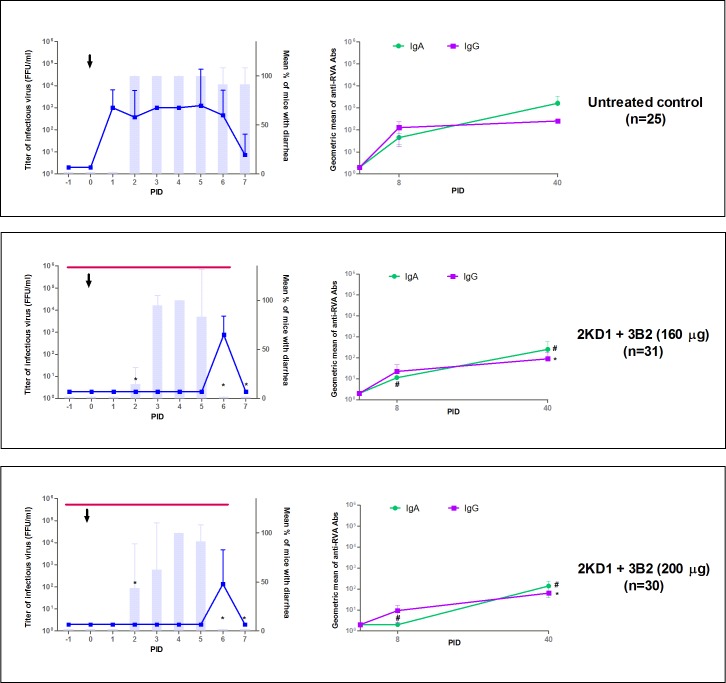
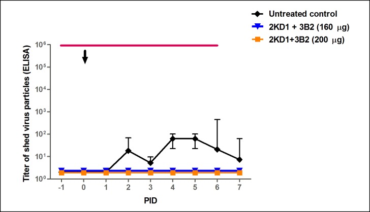
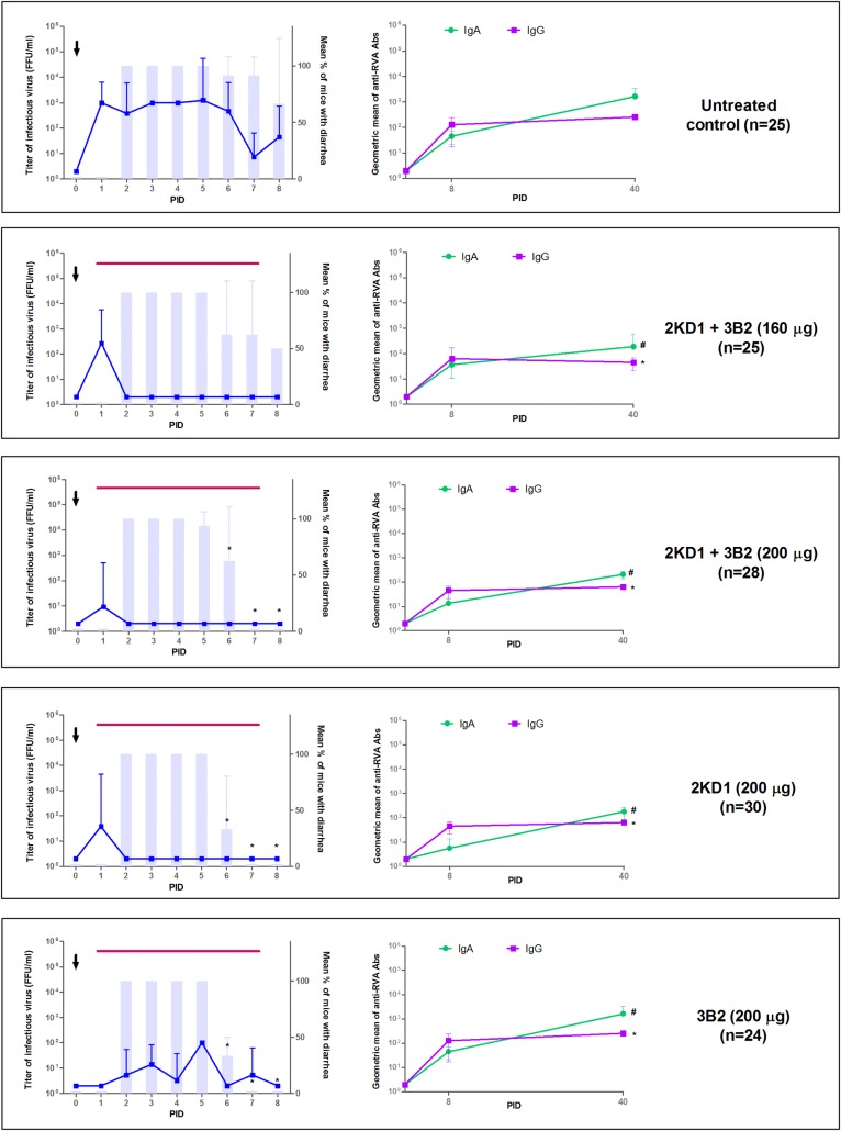
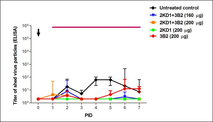
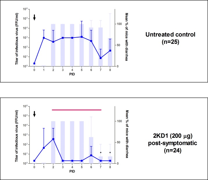
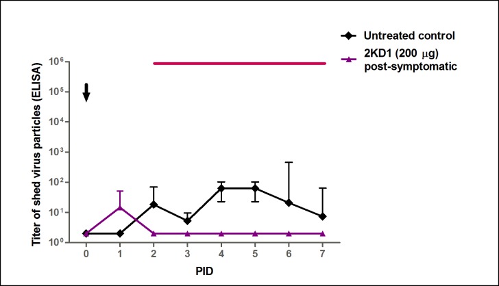
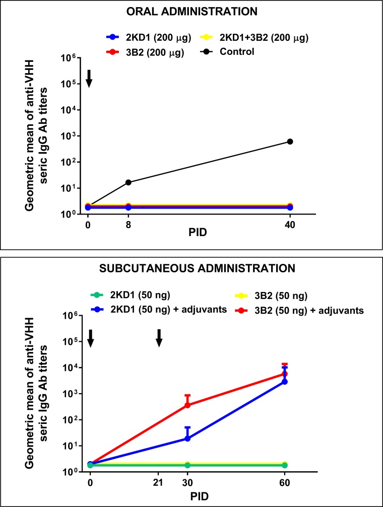
Species A Rotaviruses (RVA) remain a leading cause of mortality in children under 5 years of age. Current treatment options are limited. We assessed the efficacy of two VP6-specific llama-derived heavy chain antibody fragments (VHH) -2KD1 and 3B2- as an oral prophylactic and therapeutic treatment against RVA-induced diarrhea in a neonatal mouse model inoculated with virulent murine RVA (ECw, G16P[16]I7). Joint therapeutic administration of 2KD1+3B2 (200 μg/dose) successfully reduced diarrhea duration, RVA infection severity and virus shedding in feces. While the same dose of 2KD1 or 3B2 (200 μg) significantly reduced duration of RVA-induced diarrhea, 2KD1 was more effective in diminishing the severity of intestinal infection and RVA shedding in feces, perhaps because 2KD1 presented higher binding affinity for RVA particles than 3B2. Neither prophylactic nor therapeutic administration of the VHH interfered with the host's humoral immune response against RVA. When 2KD1 (200 μg) was administered after diarrhea development, it also significantly reduced RVA intestinal infection and fecal shedding. Host antibody responses against the oral VHH treatment were not detected, nor did viral escape mutants. Our findings show that oral administration of anti-VP6 VHH constitute, not only an effective prophylactic treatment against RVA-associated diarrhea, but also a safe therapeutic tool against RVA infection, even once diarrhea is present. Anti-VP6 VHH could be used complementary to ongoing vaccination, especially in populations that have shown lower immunization efficacy. These VHH could also be scaled-up to develop pediatric medication or functional food like infant milk formulas that might help treat RVA diarrhea.


Conflict of interest statement

The authors have declared that no competing interests exist.

Figures

Fig 1
Fig 1. Experimental design.
The thin horizontal lines represent the duration of the treatment with VHH. On PID 0, all groups were inoculated with 1778 DD50 of murine RVA (ECw). Prophylactic groups received the first daily doses of VHH on PID -1 and on PID 0, two hours before they were challenged with the viral inoculum. Pre-symptomatic and post-symptomatic treatments began on PID 1 and PID 2. Final euthanasia was performed at PID 7 for prophylactic groups and at PID 8 for therapeutic pre- and post-symptomatic groups. PID: Post Inoculation Day.
Fig 2
Fig 2. Effect of prophylactic administration of 2KD1+3B2 VHH on RVA-induced diarrhea and host humoral immune response against RVA.
All mice were orally inoculated with 1778 DD50 of ECw RVA at day four of life (PID 0), as indicated by the arrow. Mice were euthanized sequentially to obtain intestinal tissue (3–4 mice per time point). Graphics on the left side of the panels depict intestinal infectious RVA titers (by CCIF) and diarrhea prevalence. Blue lines show the development of intestinal RVA disease whereas vertical bars show the mean percentage of mice with diarrhea per group. The * symbol indicates that the percentage of mice with diarrhea was significantly lower than in the untreated control group (Fisher Exact test). The thin horizontal line indicates the duration of VHH passive treatment. Graphics on the right side of the panel display humoral immune responses against RVA. Anti-RVA IgA in intestinal samples and serum IgG Ab titers were determined by ELISA at PIDs 0, 8 and 40. The # and * symbols indicate that the Ab titers of intestinal IgA and serum IgG, respectively, were significantly lower than the corresponding Ab titers in the untreated control group (One-way ANOVA). PID: Post Inoculation day.
Fig 3
Fig 3. Effect of prophylactic administration of 2KD1+3B2 VHH on RVA shedding in feces.
Mean titer of virus shed (determined by ELISA) detected daily in pooled feces for mice treated prophylactically with VHH and the untreated control group. Treated groups did not display detectable fecal shedding at any time. The arrow shows inoculation time (PID 0) and the thin horizontal line indicates the duration of VHH passive treatment. PID: Post Inoculation Day.
Fig 4
Fig 4. Effect of pre-symptomatic therapeutic administration of VHH on RVA-induced diarrhea and host humoral immune response against RVA.
All mice were orally inoculated with ECw RVA (1778 DD50) at day four of life (PID 0), as indicated by the arrow. Mice were euthanized sequentially to obtain intestinal tissue (3–4 mice per time point). Graphics on the left side of the panel depict intestinal infectious RVA titer and the incidence of diarrhea. Blue lines show the development of intestinal RVA disease whereas vertical bars show the mean percentage of mice with diarrhea per group. The * symbol indicates that the percentage of mice with diarrhea was significantly lower than in the untreated control group (Fisher Exact test). The thin horizontal purple line indicates the duration of VHH passive treatment. Graphics on the right side of the panel display mice humoral immune response against RVA. Anti-RVA IgA in intestinal samples and serum IgG Ab titers were determined by ELISA at PID 0, 8 and 40. The # and * symbols indicate that Ab titers of intestinal IgA and serum IgG were significantly lower than Ab titers in the untreated control group (One-way ANOVA). PID: Post Inoculation Day.
Fig 5
Fig 5. Effect of pre-symptomatic therapeutic administration of 2KD1+3B2 VHH on RVA shedding in feces.
Mean titer of virus shed (determined by ELISA) detected daily in pooled feces for mice receiving pre-symptomatic therapeutic passive treatment with VHH and the untreated control group. The arrow shows virus inoculation time (PID 0) and the thin horizontal line indicates the duration of VHH passive treatment. AUC comparison showed that all VHH-treated groups, except for the ones receiving 200 μg of 3B2, significantly diminished overall fecal shedding with respect to the untreated control (ANOVA, p = 0.04). PID: Post Inoculation Day.
Fig 6
Fig 6. Effect of post-symptomatic therapeutic administration of VHH on RVA-associated diarrhea in neonatal mice.
All mice were orally inoculated with ECw RVA at day four of life (PID 0), as indicated by the arrow. Mice were euthanized sequentially to obtain intestinal tissue (3–4 mice per time point). Blue lines depict the development of intestinal RVA disease while vertical bars show the mean percentage of mice with diarrhea per group. The * symbol indicates that the percentage of mice with diarrhea was significantly lower than in the untreated control group (Fisher Exact test). The thin horizontal line shows the duration of the passive treatment. PID: Post Inoculation Day.
Fig 7
Fig 7. Effect of post-symptomatic therapeutic administration of anti-VP6 VHH on RVA shedding in feces.
Mean titer of virus shed (determined by ELISA) detected daily in pooled feces from mice receiving pre-symptomatic and post-symptomatic treatment comprising 200 μg of 2KD1 and the untreated control group. The arrow shows inoculation time (PID 0). Post-symptomatic administration of 2KD1 (200 μg) significantly reduced overall fecal shedding with respect to the untreated control group when AUC comparison was performed (ANOVA, p = 0.002). The thin horizontal line indicates the duration of VHH passive treatment. PID: Post Inoculation Day.
Fig 8
Fig 8. Humoral Ab responses against anti-RVA VHH in mice treated with anti-VP6 VHH.
The panel above shows seric IgG Ab response against anti-VP6 VHH in neonatal mice that received oral therapeutic treatment for seven days, consisting of: 2KD1 (200 μg), 3B2 (200 μg) and 2KD1+3B2 (200 μg). The control employed for this assay was serum from an adult mice immunized subcutaneously with 2KD1 (50 ng) in the presence of adjuvants. The panel below shows seric IgG Ab response against 2KD1/3B2 in adult mice immunized subcutaneously with 50 ng of VHH at PID 0 and 21 with or without adjuvants. For this experiment, four adult mice per group were used. The adjuvant employed was Freund’s complete adjuvant for the first immunization, while Freund´s incomplete adjuvant was used in the second one. Arrows indicates immunization time for both experiments.
Fig 9
Fig 9. Phylogenetic analysis of VP6 sequences.
The phylogenetic tree was constructed using the Maximum Likelihood method based on the Tamura 3-parameter model. Bootstrap statistical support (1000 repetitions) is shown. All the used strains are named with the Genbank accession number and strain name. The experimental samples are indicates as: blue circle = Prophylactic PID 7; red circle = therapeutic PIDs; black circle = untreated and; white circle = viral inoculum.
Fig 10
Fig 10. Ph stability and proteolytic resistance of anti-VP6 VHH.
The graphs show the percentage of remaining functional 2KD1/3B2 after incubation in SGF (pepsin 0.32% w/v; pH 1.2) or SIF (pancreatin 1%, w/v; pH 6.8) in different conditions, determined by RVA VHH Ab titer ELISA. The panel above shows pH stability and proteolytic for 2KD1 clone whereas the panel below depicts these parameters for 3B2. In all cases 400 μg of the VHH were incubated for one hour at 37°C. Experiments were conducted by triplicate. SGF: Simulated Gastric Fluid. SIF: Simulated Intestinal Fluid. ORS: Oral Rehydration Salts.
Fig 11
Fig 11. Binding affinity of anti-VP6 VHH to RVA particles.
Decreasing concentrations of both 2KD1 and 3B2 were tested for their binding affinity to immobilized RVA particles in an ELISA previously described. Dose-response curves were fitted using the dose-response sigmoidal model in GraphPad 7 software (available online) obtaining R square values of 0.990 for 2KD1 and 0.991 for 3B2. The EC50 values were calculated for both clones, showing that 2KD1 had a three-fold higher binding affinity for RVA particles than 3B2: 0.0016 μg/ml vs 0.0048 μg/ml.

Similar articles

Cited by

References

    1. Tate JE, Burton AH, Boschi-Pinto C, Parashar UD. Global, Regional, and National Estimates of Rotavirus Mortality in Children <5 Years of Age, 2000–2013. Clin Infect Dis. 2016;62: S96–S105. 10.1093/cid/civ1013 - DOI - PMC - PubMed
    1. WHO- UNICEF, Johns Hopkins School of Public Health U. Implementing the New Recommendations on the Clinical Management of Diarrhoea Guidelines for Policy Makers and Programme Managers. World Health Organization, Department of Child and Adolescent Health and Development, and United Nations Children’s Fund, Programme Division; 2006.
    1. Grandy G, Medina M, Soria R, Terán CG, Araya M. Probiotics in the treatment of acute rotavirus diarrhoea. A randomized, double-blind, controlled trial using two different probiotic preparations in Bolivian children. BMC Infect Dis. 2010;10: 253 10.1186/1471-2334-10-253 - DOI - PMC - PubMed
    1. Preidis GA, Saulnier DM, Blutt SE, Mistretta T, Riehle KP, Major AM, et al. Host response to probiotics determined by nutritional status of rotavirus-infected neonatal mice. J Pediatr Gastroenterol Nutr. 2012;55: 299–307. 10.1097/MPG.0b013e31824d2548 - DOI - PMC - PubMed
    1. Zhang Z, Xiang Y, Li N, Wang B, Ai H, Wang X, et al. Protective effects of Lactobacillus rhamnosus GG against human rotavirus-induced diarrhoea in a neonatal mouse model. Pathog Dis. 2013;67: 184–91. 10.1111/2049-632X.12030 - DOI - PubMed

MeSH terms

Substances