A challenging diagnosis of late-onset tumefactive multiple sclerosis associated to cervicodorsal syringomyelia: doubtful CT, MRI, and bioptic findings: Case report and literature review
- PMID: 27603348
- PMCID: PMC5023870
- DOI: 10.1097/MD.0000000000004585
A challenging diagnosis of late-onset tumefactive multiple sclerosis associated to cervicodorsal syringomyelia: doubtful CT, MRI, and bioptic findings: Case report and literature review
Abstract
Background: Tumefactive multiple sclerosis (MS) is an unusual variant of demyelinating disease characterized by lesions with pseudotumoral appearance on radiological imaging mimicking other space-occupying lesions, such as neoplasms, infections, and infarction. Especially when the patient's medical history is incompatible with MS, the differential diagnosis between these lesions constitutes a diagnostic challenge often requiring histological investigation. An older age at onset makes distinguishing tumefactive demyelinating lesion (TDL) from tumors even more challenging.
Methods: We report a case of brain TDL as the initial manifestation of late-onset MS associated with cervico-dorsal syringomyelia. A 66-year-old Caucasian woman with a 15-day history headache was referred to our hospital because of the acute onset of paraphasia. She suffered from noncommunicating syringomyelia associated to basilar impression and she reported a 10-year history of burning dysesthesia of the left side of the chest extended to the internipple line level.
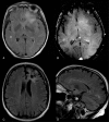
Results: Computed tomography (CT) and magnetic resonance imaging (MRI) examinations revealed a left frontal lesion with features suspicious for a tumor. Given the degree of overlap with other pathologic processes, CT and MRI findings failed to provide an unambiguous diagnosis; furthermore, because of the negative cerebrospinal fluid analysis for oligoclonal bands, the absence of other lesions, and the heightened suspicion of neoplasia, the clinicians opted to perform a stereotactic biopsy. Brain specimen analysis did not exclude the possibility of perilesional reactive gliosis and the patient, receiving anitiedemigen therapy, was monthly followed up. In the meanwhile, the second histological opinion of the brain specimen described the absence of pleomorphic glial cells indicating a tumor. These findings were interpreted as destructive inflammatory demyelinating disease and according to the evolution of MRI lesion burden, MS was diagnosed.
Conclusion: TDL still remains a problematic entity clinically, radiologically, and sometimes even pathologically. A staged follow-up is necessary, and in our case, it revealed to be the most important attitude to define the nature of the lesion, confirming the classic MS diagnostic criteria of disseminate lesions in time and space. We discuss our findings according to the recent literature.
Conflict of interest statement
The authors have no funding and conflicts of interest to disclose.
Figures


References
Publication types
MeSH terms
LinkOut - more resources
Full Text Sources
Other Literature Sources
Medical

