

Use of Digitally Stained Multimodal Confocal Mosaic Images to Screen for Nonmelanoma Skin Cancer
- PMID: 27603676
- PMCID: PMC5757842
- DOI: 10.1001/jamadermatol.2016.2997
Use of Digitally Stained Multimodal Confocal Mosaic Images to Screen for Nonmelanoma Skin Cancer
Abstract
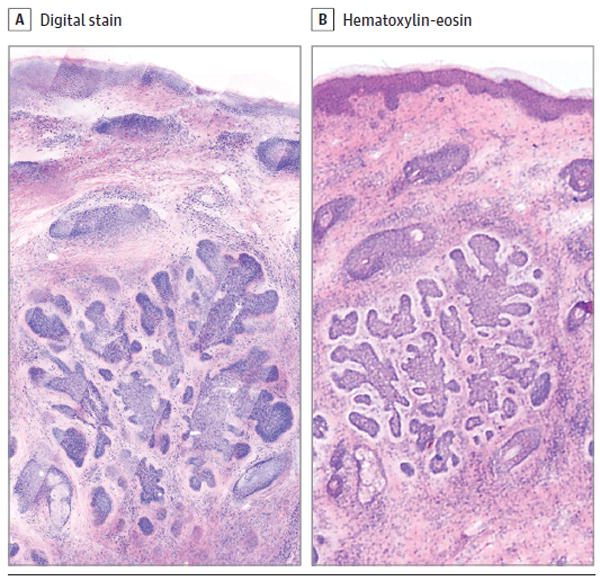
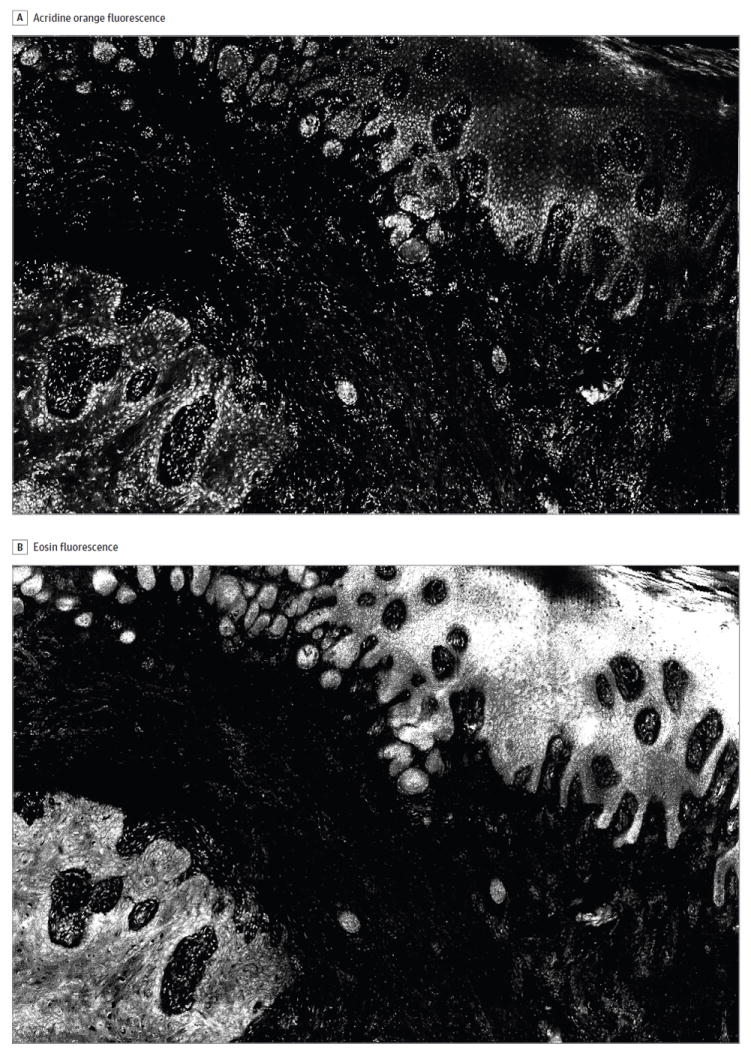
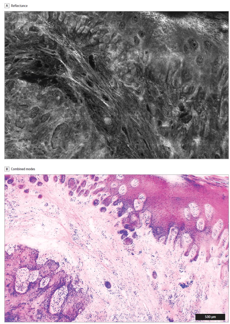
Importance: Confocal microscopy has the potential to provide rapid bedside pathologic analysis, but clinical adoption has been limited in part by the need for physician retraining to interpret grayscale images. Digitally stained confocal mosaics (DSCMs) mimic the colors of routine histologic specimens and may increase adaptability of this technology.
Objective: To evaluate the accuracy and precision of 3 physicians using DSCMs before and after training to detect basal cell carcinoma (BCC) and squamous cell carcinoma (SCC) in Mohs micrographic surgery fresh-tissue specimens.
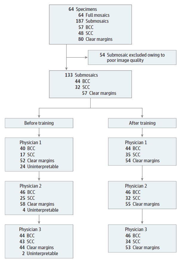
Design: This retrospective study used 133 DSCMs from 64 Mohs tissue excisions, which included clear margins, residual BCC, or residual SCC. Discarded tissue from Mohs surgical excisions from the dermatologic surgery units at Memorial Sloan Kettering Cancer Center and Oregon Health & Science University were collected for confocal imaging from 2006 to 2011. Final data analysis and interpretation took place between 2014 and 2016. Two Mohs surgeons and a Mohs fellow, who were blinded to the correlating gold standard frozen section diagnoses, independently reviewed the DSCMs for residual nonmelanoma skin cancer (NMSC) before and after a brief training session (about 5 minutes). The 2 assessments were separated by a 6-month washout period.
Main outcomes and measures: Diagnostic accuracy was characterized by sensitivity and specificity of detecting NMSC using DSCMs vs standard frozen histopathologic specimens. The diagnostic precision was calculated based on interobserver agreement and κ scores. Paired 2-sample t tests were used for comparative means analyses before and after training.
Results: The average respective sensitivities and specificities of detecting NMSC were 90% (95% CI, 89%-91%) and 79% (95% CI, 52%-100%) before training and 99% (95% CI, 99%-99%) (P = .001) and 93% (95% CI, 90%-96%) (P = .18) after training; for BCC, they were 83% (95% CI, 59%-100%) and 92% (95% CI, 81%-100%) before training and 98% (95% CI, 98%-98%) (P = .18) and 97% (95% CI, 95%-100%) (P = .15) after training; for SCC, they were 73% (95% CI, 65%-81%) and 89% (95% CI, 72%-100%) before training and 100% (P = .004) and 98% (95% CI, 95%-100%) (P = .21) after training. The pretraining interobserver agreement was 72% (κ = 0.58), and the posttraining interobserver agreement was 98% (κ = 0.97) (P = .04).
Conclusions and relevance: Diagnostic use of DSCMs shows promising correlation to frozen histologic analysis, but image quality was affected by variations in image contrast and mosaic-stitching artifact. With training, physicians were able to read DSCMs with significantly improved accuracy and precision to detect NMSC.
Conflict of interest statement
Figures
Similar articles
-
Detection of basal cell carcinomas in Mohs excisions with fluorescence confocal mosaicing microscopy.Br J Dermatol. 2009 Jun;160(6):1242-50. doi: 10.1111/j.1365-2133.2009.09141.x. Epub 2009 Mar 30. Br J Dermatol. 2009. PMID: 19416248 Free PMC article.
-
Diagnostic accuracy of autofluorescence-Raman microspectroscopy for surgical margin assessment during Mohs micrographic surgery of basal cell carcinoma.Br J Dermatol. 2024 Aug 14;191(3):428-436. doi: 10.1093/bjd/ljae196. Br J Dermatol. 2024. PMID: 38736216
-
Detection of skin cancer margins in Mohs excisions with high-speed strip mosaicing confocal microscopy: a feasibility study.Br J Dermatol. 2013 Oct;169(4):922-6. doi: 10.1111/bjd.12444. Br J Dermatol. 2013. PMID: 23701464 Free PMC article.
-
Surgical margins in the treatment of nonmelanoma skin cancer and mohs micrographic surgery.Curr Surg. 2005 Sep-Oct;62(5):518-26. doi: 10.1016/j.cursur.2005.01.003. Curr Surg. 2005. PMID: 16125611 Review.
-
Upstaging of basal cell and squamous cell carcinomas during definitive surgery: a review of predictive preoperative clinical and histologic features.Arch Dermatol Res. 2021 Jul;313(5):319-325. doi: 10.1007/s00403-020-02151-5. Epub 2020 Oct 27. Arch Dermatol Res. 2021. PMID: 33108525 Review.
Cited by
-
Deep learning automated pathology in ex vivo microscopy.Biomed Opt Express. 2021 May 5;12(6):3103-3116. doi: 10.1364/BOE.422168. eCollection 2021 Jun 1. Biomed Opt Express. 2021. PMID: 34221648 Free PMC article.
-
Updates on the Management of Non-Melanoma Skin Cancer (NMSC).Healthcare (Basel). 2017 Nov 1;5(4):82. doi: 10.3390/healthcare5040082. Healthcare (Basel). 2017. PMID: 29104226 Free PMC article. Review.
-
An international 3-center training and reading study to assess basal cell carcinoma surgical margins with ex vivo fluorescence confocal microscopy.J Cutan Pathol. 2021 Aug;48(8):1010-1019. doi: 10.1111/cup.13980. Epub 2021 Mar 3. J Cutan Pathol. 2021. PMID: 33576022 Free PMC article.
-
Skin Imaging Using Ultrasound Imaging, Optical Coherence Tomography, Confocal Microscopy, and Two-Photon Microscopy in Cutaneous Oncology.Front Med (Lausanne). 2019 Nov 22;6:274. doi: 10.3389/fmed.2019.00274. eCollection 2019. Front Med (Lausanne). 2019. PMID: 31824956 Free PMC article. Review.
-
Line scanning, stage scanning confocal microscope (LSSSCM).Biomed Opt Express. 2017 Jul 24;8(8):3807-3815. doi: 10.1364/BOE.8.003807. eCollection 2017 Aug 1. Biomed Opt Express. 2017. PMID: 28856051 Free PMC article.
References
-
- Rogers HW, Weinstock MA, Feldman SR, Coldiron BM. Incidence estimate of nonmelanoma skin cancer (keratinocyte carcinomas) in the US population. JAMA Dermatol. 2012;151(10):1081–1086. - PubMed
-
- Rajadhyaksha M, Grossman M, Esterowitz D, Webb RH, Anderson RR. In vivo confocal scanning laser microscopy of human skin: melanin provides strong contrast. J Invest Dermatol. 1995;104(6):946–952. - PubMed
-
- Ulrich M, Lange-Asschenfeldt S. In vivo confocal microscopy in dermatology: from research to clinical application. J Biomed Opt. 2013;18(6):061212. - PubMed
-
- New KC, Petroll WM, Boyde A, et al. In vivo imaging of human teeth and skin using real-time confocal microscopy. Scanning. 1991;13(5):369–372.
-
- González S. Confocal reflectance microscopy in dermatology: promise and reality of non-invasive diagnosis and monitoring. Actas Dermosifiliogr. 2009;100(suppl 2):59–69. - PubMed
Publication types
MeSH terms
Grants and funding
LinkOut - more resources
Full Text Sources
Other Literature Sources
Medical
Molecular Biology Databases
Research Materials
