Case Reports
. 2016 Sep 7;16(1):131.
doi: 10.1186/s12890-016-0293-2.

Dental technician pneumoconiosis mimicking pulmonary tuberculosis: a case report

Affiliations
Case Reports

Dental technician pneumoconiosis mimicking pulmonary tuberculosis: a case report

Han Loong Tan et al. BMC Pulm Med. .

Abstract

Background: Dental laboratory technicians are at risk of developing occupational respiratory diseases due to exposure to various potentially toxic substances in their working environment. Since 1939, few cases of silicosis among dental technician have been reported.

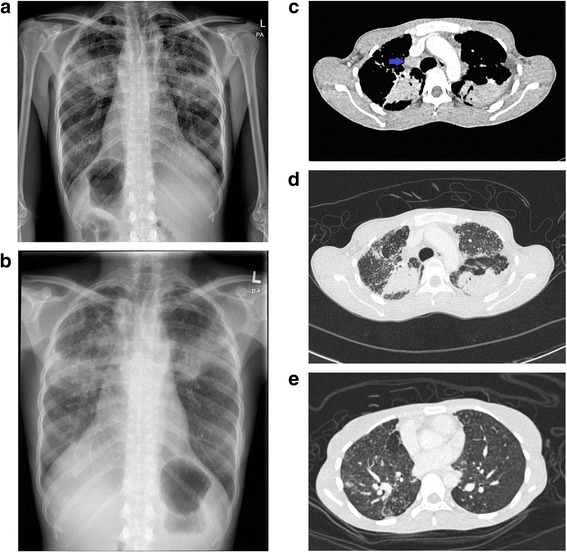
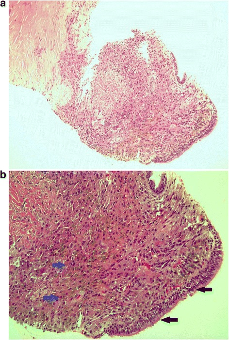
Case presentation: We illustrate a 38 year-old female, who worked in a dental laboratory for 20 years, initially treated as pulmonary tuberculosis and chronic necrotising aspergillosis without much improvement. Computed tomography guided lung biopsy and bronchoscopic transbronchial lung biopsy were performed. Lung tissue biopsies showed presence of refractile dental materials within the areas of histiocyte proliferation. The diagnosis of dental technician pneumoconiosis was obtained and our patient underwent pulmonary rehabilitation.

Conclusions: This case highlights the importance of obtaining a detailed occupational history in tuberculosis endemic area, as pulmonary tuberculosis is a great mimicker of other respiratory diseases.

Keywords: Case report; Dental technician pneumoconiosis; Pulmonary tuberculosis.


Figures

Fig. 1
Fig. 1
a Chest x-ray showed fibrotic changes at both upper zones with reticulo-nodular opacity over the upper and middle zone of both lungs. b Repeated chest x-ray after 54 doses of anti tuberculous medications showed absence of radiological improvement with similar reticulo-nodular opacity over bilateral lung field. c Mediastinal view of CT thorax showed enlarged right paratrachea lymphadenopathy (Blue arrow). d Consolidative changes with air bronchogram at the posterior segment of the left upper lobe and the apico-posterior segment of the right upper lobe. e The presence of lung nodules with tree in bud appearance
Fig. 2
Fig. 2
a and b CT guided lung biopsy. Haematoxylin and eosin stain revealed absence of normal alveolar tissue, which were replaced by fibrous tissue. There were areas of histiocytes proliferation with the presence of refractile dental material (blue arrow). The fibrous tissue was lined by ciliated tall columnar respiratory epithelium (black arrow)

Similar articles

Cited by

References

    1. Seldén A, Sahle W, Johansson L, Sörenson S, Persson B. Three Cases of Dental Technician’s Pneumoconiosis Related to Cobalt-Chromium-Molybdenum Dust Exposure. Chest. 1996;109(3):837–42. doi: 10.1378/chest.109.3.837. - DOI - PubMed
    1. Rom W, Lockey J, Lee J, et al. Pneumoconiosis and exposures of dental laboratory technicians. Am J Public Health. 1984;74(11):1252–7. doi: 10.2105/AJPH.74.11.1252. - DOI - PMC - PubMed
    1. Selden A, Persson B, Bornberger-Dankvardt S, Winstrom L, Bodin L. Exposure to cobalt chromium dust and lung disorders in dental technicians. Thorax. 1995;50(7):769–72. doi: 10.1136/thx.50.7.769. - DOI - PMC - PubMed
    1. Dogan DO, Ozdemir AK, Polat NT, Dal U, Gumus C, Akkurt I. Prevalence of respiratory abnormalities and pneumoconiosis in dental laboratory technicians. Tuberk Toraks. 2010;58:135–41. - PubMed
    1. Berk S, Dogan D, Gumus C, Akkurt I. Relationship between radiological (X-ray/HRCT), spirometric and clinical findings in dental technicians’ pneumoconiosis. Clin Respir J. 2014;10(1):67–73. doi: 10.1111/crj.12187. - DOI - PubMed

Publication types