. 2017 Jan;61(1):42-51.
doi: 10.1007/s12031-016-0830-8. Epub 2016 Sep 7.

Beneficial Effects of a CaMKIIα Inhibitor TatCN21 Peptide in Global Cerebral Ischemia

Affiliations

Beneficial Effects of a CaMKIIα Inhibitor TatCN21 Peptide in Global Cerebral Ischemia

Mohammad Ejaz Ahmed et al. J Mol Neurosci. 2017 Jan.

Abstract

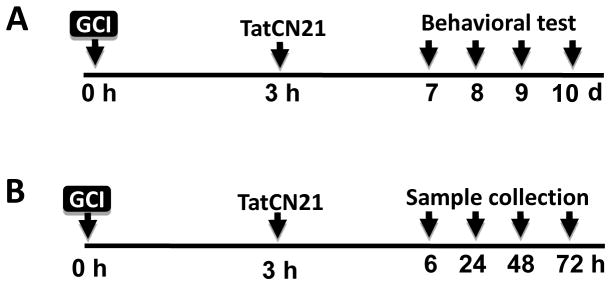
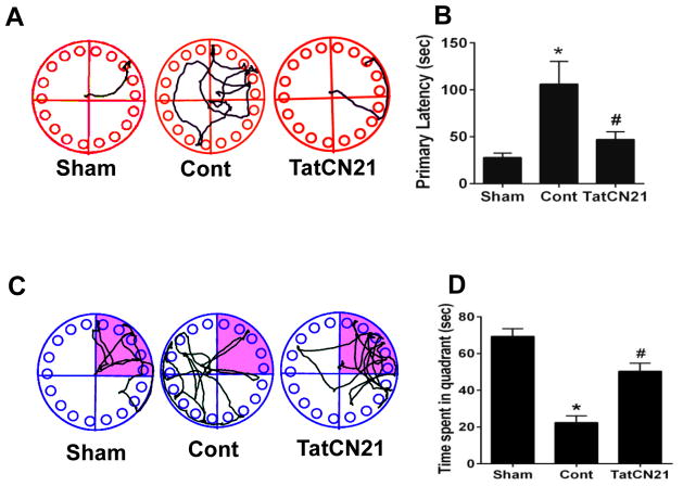
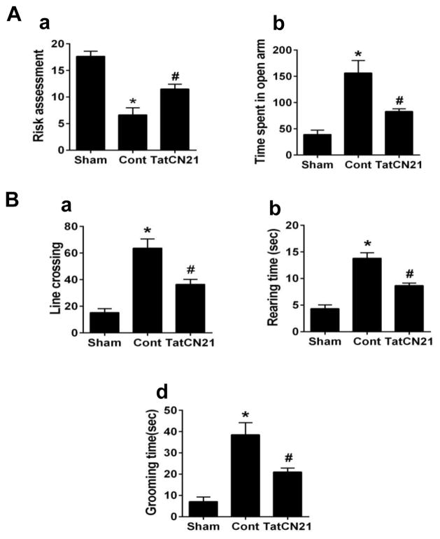
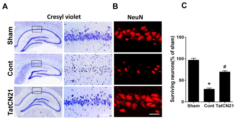
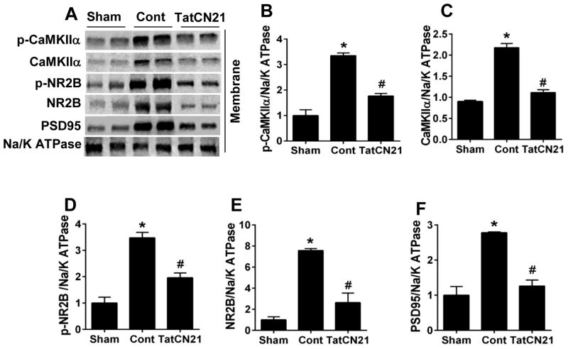
Aberrant calcium influx is a common feature following ischemic reperfusion (I/R) in transient global cerebral ischemia (GCI) and causes delayed neuronal cell death in the CA1 region of the hippocampus. Activation of calcium-calmodulin (CaM)-dependent protein kinase IIα (CaMKIIα) is a key event in calcium signaling in ischemic injury. The present study examined the effects of intracerebroventricular (icv) injection of tatCN21 in ischemic rats 3 h after GCI reperfusion. Cresyl violet and NeuN staining revealed that tatCN21 exerted neuroprotective effects against delayed neuronal cell death of hippocampal CA1 pyramidal neurons 10 days post-GCI. In addition, TatCN21 administration ameliorated GCI-induced spatial memory deficits in the Barnes maze task as well as anxiety-like behaviors and spontaneous motor activity in the elevated plus maze and open field test, respectively. Mechanistic studies showed that the administration of tatCN21 decreased GCI-induced phosphorylation, translocation, and membrane targeting of CaMKIIα. Treatment with tatCN21 also inhibited the level of CaMKIIα-NR2B interaction and NR2B phosphorylation. Our results revealed an important role of tatCN21 in inhibiting CaMKIIα activation and its beneficial effects in neuroprotection and memory preservation in an ischemic brain injury model.

Keywords: CaMKIIα; Global cerebral ischemia; Hippocampus; NR2B; TatCN21.


Conflict of interest statement

The author declares that there is no conflict of interest.

Figures

Fig. 1
Fig. 1
Schematic diagram depicting experimental design. (A) GCI model was developed at 0 h, and tatCN21 (50 μg/rat) was icv injected 3 h after GCI. On day 7 through day 10, behavioral tests were performed to check learning and memory, anxiety, and spontaneous motor activity. Rats were anesthetized with sodium pentobarbital, transcardially perfused and sacrificed on day 10 and brain sections were collected. (B) 3 h after GCI, animals were treated with tatCN21 and scrambled tat peptide (cont) and sacrificed at 6, 24, 48 and 72 h after reperfusion. Whole brains were quickly removed and hippocampal CA1 region were rapidly micro dissected, immediately frozen and preserved for different assay.
Fig. 2
Fig. 2
Icv infusion of tatCN21 peptide 3 h post GCI significantly attenuates the hippocampal dependent learning and memory deficits following GCI. (A, B) Barnes maze test was performed to check the spatial learning and memory ability of sham, control (cont) and tatCN21 group animals, as shown by latency to find the black escape box on day 9 following GCI. (C, D) Probe trials measure the time spent in the quadrant where the escape box was previously located but removed on day 10 after GCI. The representative tracks are shown and data were statistically compared. Data are represented as mean ±SE (n=10) per group, *P <0.05 vs. sham, #P < 0.05 vs. cont group.
Fig. 3
Fig. 3
TatCN21 infusion 3 h after GCI reduced anxiety and locomotor activity behaviors in the elevated plus maze and open field test. (A) In the elevated plus maze test, the number of risk assessment and time spent in open arm were calculated and statistically compared. (B) In the open field test, number of line crossed, rearing, grooming were recorded and statistically analyzed. Data are represented as mean ±SE (n=10) per group, *P <0.05 vs. sham, #P < 0.05 vs. cont group.
Fig. 4
Fig. 4
TatCN21 confers neuroprotection in hippocampal CA1 region on day 10 after GCI. (A) Cresyl violet staining shows the whole hippocampus overview and the CA1 region. Bar scale: 50 μm. (B) High magnification immunofluorescence staining of NeuN shows pyramidal neurons in the medial hippocampal CA1 region. Bar scale: 20 μm. (C) The number of surviving neurons was quantified and statistically compared. Data were represented as mean ± SE (n=6) per group, *P <0.05 vs. sham, #P <0.05 vs. cont group.
Fig. 5
Fig. 5
Effect of tatCN21 on the phosphorylation of CaMKIIα, NR2B and their interactions in the hippocampal CA1 region 6 h after GCI. (A, B) Time course study of the cytosolic phosphorylation levels of CaMKIIα and NR2B at 6, 24, 48 and 72 h post GCI. Bands were scanned and the levels of p-CaMKIIα/CaMKIIα and p-NR2B/NR2B were determined. (C, D) Immunoblotting analysis of the p-CaMKIIα/CaMKIIα in the cytosolic fraction 6 h after GCI. (E) Representative immunofluorescence staining of p-CaMKIIα and p-NR2B in the CA1 region of hippocampus. Bar scale: 20 μm. Data are represented as mean ± SE (n=6) per group, *P <0.05 vs. sham, #P < 0.05 vs. cont group.
Fig. 6
Fig. 6
TatCN21 reduced the membrane translocation of CaMKIIα, NR2B and PSD95 in hippocampal CA1 region 6 h after GCI. (A) Representative western blots of the indicated proteins using membrane proteins at 6 h after GCI. (B–F) Normalized protein expression levels of p-CaMKIIα, CaMKIIα, p-NR2B, NR2B and PSD95 were quantified and statistically compared. Data are represented as mean ± SE (n=6) per group, *P <0.05 vs. sham, #P < 0.05 vs. cont group.
Fig. 7
Fig. 7
TatCN21 peptide decreased the interaction of CaMKIIα with NR2B following 6 h I/R. (A) Representative Co-IP of CaMKIIα and NR2B protein sample were immunoprecipated with NR2B antibody and WB with anti-CaMKIIα antibody. A reversal of Co-IP was also performed. (B, C) The binding level of CaMKIIα and NR2B were analyzed and shown as fold increased vs. Sham. (D) Duolink II in situ Proximity ligation assay using CaMKII α and NR2B antibodies in CA1 region of hippocampus 6 h after GCI. For clarity, both color and black and white images of Duolink puncta are provided. Bar scale: 50 μm. (E) Quantification of the Duolink puncta confirms that tatCN21 peptide treatment significantly reduces interaction of CaMKIIα and NR2B in the hippocampal CA1 region 6 h after GCI, compared with cont group. Data are represented as mean ± SE (n=6) per group, *P <0.05 vs. sham, #P < 0.05 vs. cont group, magnification 40x.

References

    1. Aronowski J, Grotta JC, Waxham MN. Ischemia-induced translocation of Ca2+/calmodulin-dependent protein kinase II: potential role in neuronal damage. Journal of neurochemistry. 1992;58:1743–1753. - PubMed
    1. Ashpole NM, Hudmon A. Excitotoxic neuroprotection and vulnerability with CaMKII inhibition. Molecular and cellular neurosciences. 2011;46:720–730. - PubMed
    1. Babcock AM, Everingham A, Paden CM, Kimura M. Baclofen is neuroprotective and prevents loss of calcium/calmodulin-dependent protein kinase II immunoreactivity in the ischemic gerbil hippocampus. Journal of neuroscience research. 2002;67:804–811. - PubMed
    1. Barnes CA. Memory deficits associated with senescence: a neurophysiological and behavioral study in the rat. Journal of comparative and physiological psychology. 1979;93:74–104. - PubMed
    1. Bothe HW, Bosma HJ, Hofer H, Hossmann KA, Angermeier WF. Selective vulnerability of hippocampus and disturbances of memory storage after mild unilateral ischemia of gerbil brain. Stroke; a journal of cerebral circulation. 1986;17:1160–1163. - PubMed

MeSH terms