. 2016 Sep 8:6:32956.
doi: 10.1038/srep32956.

Mapping the complete glycoproteome of virion-derived HIV-1 gp120 provides insights into broadly neutralizing antibody binding

Affiliations

Mapping the complete glycoproteome of virion-derived HIV-1 gp120 provides insights into broadly neutralizing antibody binding

Maria Panico et al. Sci Rep. .

Abstract

The surface envelope glycoprotein (SU) of Human immunodeficiency virus type 1 (HIV-1), gp120(SU) plays an essential role in virus binding to target CD4+ T-cells and is a major vaccine target. Gp120 has remarkably high levels of N-linked glycosylation and there is considerable evidence that this "glycan shield" can help protect the virus from antibody-mediated neutralization. In recent years, however, it has become clear that gp120 glycosylation can also be included in the targets of recognition by some of the most potent broadly neutralizing antibodies. Knowing the site-specific glycosylation of gp120 can facilitate the rational design of glycopeptide antigens for HIV vaccine development. While most prior studies have focused on glycan analysis of recombinant forms of gp120, here we report the first systematic glycosylation site analysis of gp120 derived from virions produced by infected T lymphoid cells and show that a single site is exclusively substituted with complex glycans. These results should help guide the design of vaccine immunogens.


Figures

Figure 1
Figure 1. Virion-associated Env analysis.
(a) A two-dye SYPRO-stained (total protein magenta, glycoproteins green) SDS-PAGE gel analysis of early (1) vs. long-term culture (2,3) of HIV-1BaL. Early HIV-1BaL was prepared from a short term passage culture using virus originally obtained from the NIH AIDS Research and Reference Reagent Program. Long-term samples were derived from the original cultures, after extended culture. Gel was calibrated with the gp120SU (200–12.5 ng) and purified p24CA (600–37.5 ng) standards. MW standards are found in lane 4. Positive control samples are found in lanes 5–7; SIVmac239,(5), HIV-1NL43 (6), HIV-1MN (7). (b) Table shows calculated Gag:Env ratios for the different virus preparations based on densitometric measurements normalized for molecular weights, and estimated average trimers per virion, assuming 1400 gag molecules per particle. (c) mAb 4E10 anti-HIV-1 TM immunoblot analysis of early (2) vs. long-term (3) HIV-1BaL culture series; 1 is MW standards. Short term passage virus shows only full length TM. In contrast, long-term cultured virus shows a mixture of TM species including both full length gp41 and truncated ~gp36.
Figure 2
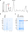
Figure 2. HPLC fractionation of HIV-1 BaL/SupT1-R5 used to purify gp120.
(a) HPLC was used to separate HIV-1 BaL/SUPT1-R5 after inactivation with aldrithiol-2 under non-reducing conditions using 206nm UV absorbance to detect proteins. Viral protein peaks (identified by SDS-PAGE gels, sequencing and immunoblot analysis) are labelled above the chromatograph. (b) Coomassie blue-stain of SDS-PAGE gel was used to analyse gp120SU corresponding fractions, with the molecular mass of standards denoted (Lane 1). Lane 2 represents pre-fractionated, long term cultured virus (1μL). We analysed HPLC fractions (Lanes 3–5). (c) Coomassie blue stained SDS-PAGE gel following HPLC purification of gp120SU (lanes 2–4). Lanes 1 represent MW standards. The band containing gp120SU was excised for glycomic/gylcoproteomic analyses.
Figure 3
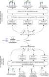
Figure 3. Simplified workflow of the glycomic and glycoproteomic methodologies employed.
The samples, in the form of isolated gp120 gel bands, were excised and proteolytically digested in-gel. The resultant peptides/glycopeptides were then eluted and subjected to either a detailed glycomic workflow (upper half of the figure) or in-depth glycoproteomic analyses (lower half of the figure).
Figure 4
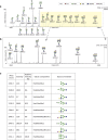
Figure 4. N-glycome of HIV-1 BaL gp120.
MALDI TOF mass spectrum of N-glycans released after PNGase-F digest: (a) full range spectrum (m/z 1500–4300); (b) expanded region (m/z 2500–4300), highlighted in the full range mass spectrum. Signal abundance (relative intensity) is normalised to the most abundant ion (indicated on the right hand axis) of the specified mass range. Structural assignments were based on MS and MS/MS data and knowledge of N-glycan biosynthetic pathways. The satellite peaks near the major peaks are permethylation artefacts whilst the peaks which are labelled with an “x” are derived from known contaminants. (c) Tabulated MALDI TOF data for N-glycans released after Endo-H digest.
Figure 5
Figure 5. Visual representation of the HIV-1BaL gp120 secondary structure.
Residue numbers for N-linked glycosylation sites are listed as both sequential numbering with respect to the experimentally deduced BaL sequence, and with the numbering with respect to the reference HIVHXB2 indicated in brackets. Individual sites are indicated as orange circles, with the sequential site number (1–24) shown within the circle. The three consensus sites of HXB2CG not present in this swarm are indicated with empty circles. The general type of glycosylation (high mannose, complex, hybrid) observed at each site is indicated by colour coding, with the key given within the figure.
Figure 6
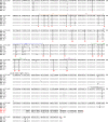
Figure 6. Complete gp160 amino acid alignment of HIV-1 BaL/SupT1-R5.
BAL.AY713409 is the published reference sequence (GenBank accession number AY713409). Dashed lines show sequence identity to the reference sequence and amino acid polymorphisms are indicated by a single letter amino acid abbreviation. Key landmarks in gp160 are denoted above each region. Potential N-linked glycosylation sites are shaded. In two sequences (H11 and E1), a nucleotide frame shift lead to a premature stop codon (*). In a third sequence (C12) a nucleotide change in W754 also led to a premature stop codon (*).

References

    1. Pejchal R. et al. A potent and broad neutralizing antibody recognizes and penetrates the HIV glycan shield. Science 334, 1097–1103, doi: 10.1126/science.1213256 (2011). - DOI - PMC - PubMed
    1. Qi Y., Jo S. & Im W. Roles of glycans in interactions between gp120 and HIV broadly neutralizing antibodies. Glycobiology 26, 251–260, doi: 10.1093/glycob/cwv101 (2016). - DOI - PMC - PubMed
    1. Mizuochi T. et al. Carbohydrate structures of the human-immunodeficiency-virus (HIV) recombinant envelope glycoprotein gp120 produced in Chinese-hamster ovary cells. Biochem J 254, 599–603 (1988). - PMC - PubMed
    1. Zhu X., Borchers C., Bienstock R. J. & Tomer K. B. Mass spectrometric characterization of the glycosylation pattern of HIV-gp120 expressed in CHO cells. Biochemistry 39, 11194–11204 (2000). - PubMed
    1. Go E. P. et al. Glycosylation site-specific analysis of clade C HIV-1 envelope proteins. Journal of proteome research 8, 4231–4242, doi: 10.1021/pr9002728 (2009). - DOI - PMC - PubMed

Publication types

MeSH terms