Case Reports
. 2016 Sep 7;10(1):246.
doi: 10.1186/s13256-016-1024-2.

Bilateral nasolabial cysts: a case report

Affiliations
Case Reports

Bilateral nasolabial cysts: a case report

Masaru Sato et al. J Med Case Rep. .

Abstract

Background: Nasoalveolar cysts are rare non-odontogenic cysts that occur beneath the nasal alar region. Few cases of bilateral nasoalveolar cysts have been described.

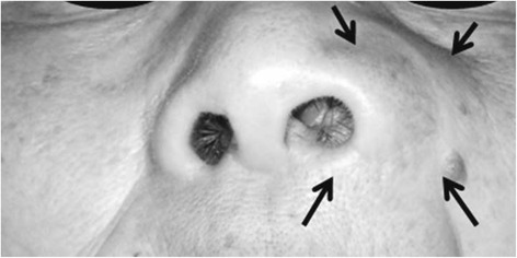
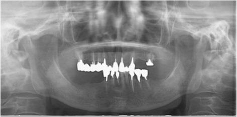
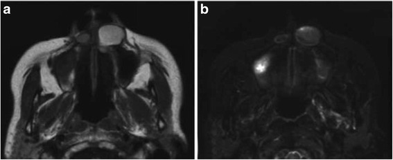
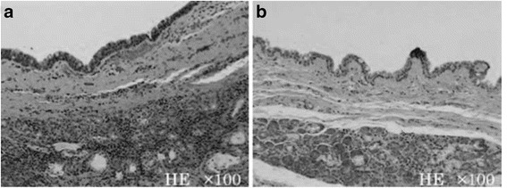
Case presentation: We report a rare case of a 67-year-old Japanese woman with bilateral nasoalveolar cysts who presented to our department with the chief complaint of a swollen left nasal alar base. Panoramic radiography revealed no abnormalities. Computed tomography and magnetic resonance imaging revealed a well-circumscribed oval lesion at both alar bases. Therefore, bilateral nasoalveolar cysts were clinically diagnosed. Furthermore, these cysts were extirpated under general anesthesia; the aforementioned diagnosis was histopathologically confirmed. No recurrence has been observed 1 year after surgery.

Conclusions: Nasoalveolar cysts are rare. It is necessary to be careful because nasoalveolar cysts can show bilateral occurrence.

Keywords: Bilateral lesions; Nasoalveolar cysts; Non-odontogenic cysts.


Figures

Fig. 1
Fig. 1
Extra oral findings of the patient at the first visit. A diffuse swelling without tenderness was found at her left nasal alar base (arrows)
Fig. 2
Fig. 2
Panoramic radiograph obtained at the first examination. Only apical periodontitis of the left upper central incisor was observed
Fig. 3
Fig. 3
Computed tomography image obtained at the first examination. A well-circumscribed oval lesion, approximately 19 × 14 × 12 mm in size, was found inferior to the left nasal alar base. A small high-density area was observed adjacent to the right piriform aperture
Fig. 4
Fig. 4
Magnetic resonance images obtained just before surgery. a. T1-weighted image. Oval lesions with high internal signal intensity inferior to the bilateral alar base. b. T2-weighted short inversion time inversion recovery image showing low signal intensity
Fig. 5
Fig. 5
Hematoxylin and eosin-stained sections of the surgical specimen. a. Right-sided lesion. b. Left-sided lesion. The cysts were lined with thin, non-keratinized squamous epithelium containing a number of goblet cells. The cyst walls comprised fibrous connective tissue with seromucous glands

References

    1. Aikawa T, Iida S, Fukuda Y, Nakano Y, Ota Y, Takao K, et al. Nasolabial cyst in a patient with cleft lip and palate. Int J Oral Maxillofac Surg. 2008;37:874–6. doi: 10.1016/j.ijom.2008.04.016. - DOI - PubMed
    1. Choi JH, Cho JH, Kang HJ, Chae SW, Lee SH, Hwang SJ, et al. Nasolabial cyst: a retrospective analysis of 18 cases. Ear Nose Throat J. 2002;81:94–6. - PubMed
    1. el-Din K, el-Hamd AA. Nasolabial cyst: a report of eight cases and a review of the literature. J Laryngol Otol. 1999;113:747–9. - PubMed
    1. Iida S, Aikawa T, Kishino M, Sakai T, Nakano Y, Okura M, et al. Spheric mass beneath the alar base: MR images of nasolabial cyst and schwannoma. AJNR Am J Neuroradiol. 2006;27:1826–9. - PMC - PubMed
    1. Kato H, Kanematsu M, Kusunoki Y, Shibata T, Murakami H, Mizuta K, et al. Nasoalveolar cyst: imaging findings in three cases. Clin Imaging. 2007;31:206–9. doi: 10.1016/j.clinimag.2006.12.026. - DOI - PubMed

Publication types