. 2016 Nov;6(11):1258-1266.
doi: 10.1158/2159-8290.CD-16-0267. Epub 2016 Sep 7.

Biallelic Alteration and Dysregulation of the Hippo Pathway in Mucinous Tubular and Spindle Cell Carcinoma of the Kidney

Affiliations

Biallelic Alteration and Dysregulation of the Hippo Pathway in Mucinous Tubular and Spindle Cell Carcinoma of the Kidney

Rohit Mehra et al. Cancer Discov. 2016 Nov.

Abstract

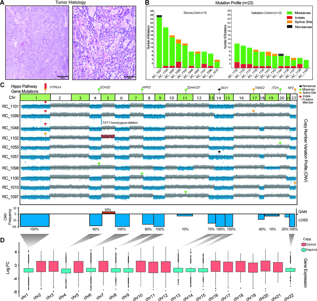
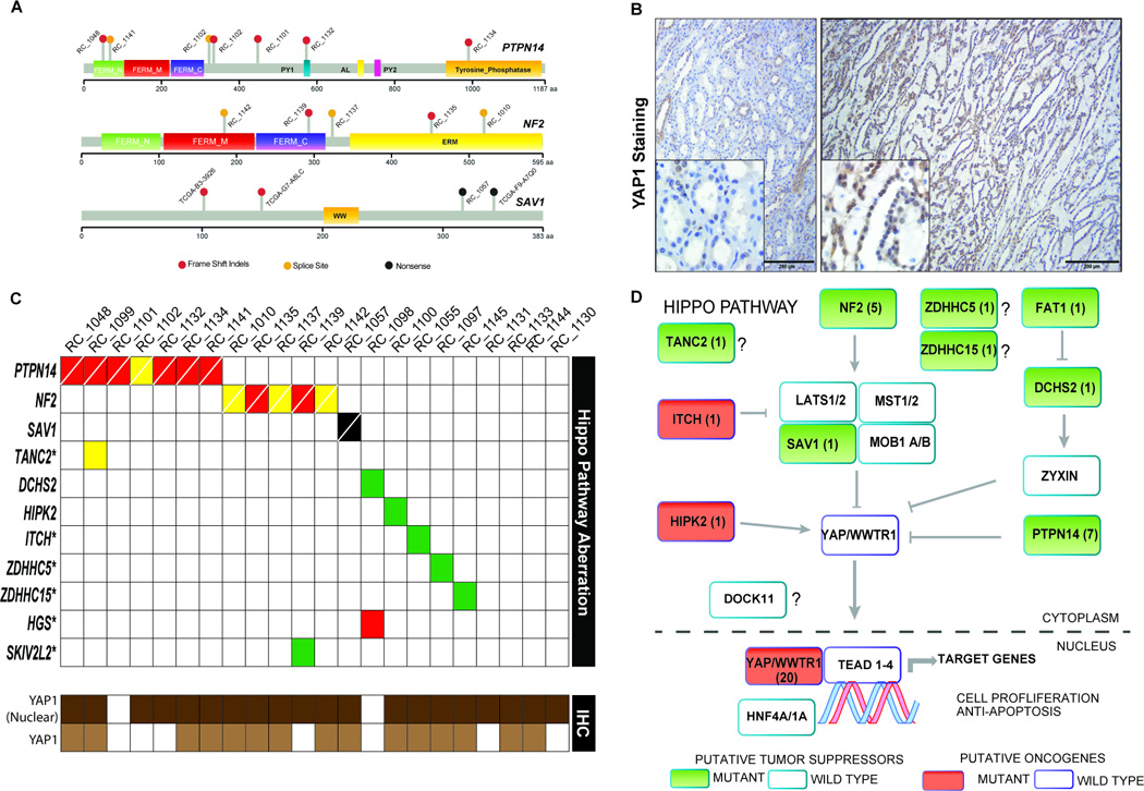
Mucinous tubular and spindle cell carcinoma (MTSCC) is a relatively rare subtype of renal cell carcinoma (RCC) with distinctive morphologic and cytogenetic features. Here, we carry out whole-exome and transcriptome sequencing of a multi-institutional cohort of MTSCC (n = 22). We demonstrate the presence of either biallelic loss of Hippo pathway tumor suppressor genes (TSG) and/or evidence of alteration of Hippo pathway genes in 85% of samples. PTPN14 (31%) and NF2 (22%) were the most commonly implicated Hippo pathway genes, whereas other genes such as SAV1 and HIPK2 were also involved in a mutually exclusive fashion. Mutations in the context of recurrent chromosomal losses amounted to biallelic alterations in these TSGs. As a readout of Hippo pathway inactivation, a majority of cases (90%) exhibited increased nuclear YAP1 protein expression. Taken together, nearly all cases of MTSCC exhibit some evidence of Hippo pathway dysregulation.

Significance: MTSCC is a rare and relatively recently described subtype of RCC. Next-generation sequencing of a multi-institutional MTSCC cohort revealed recurrent chromosomal losses and somatic mutations in the Hippo signaling pathway genes leading to potential YAP1 activation. In virtually all cases of MTSCC, there was evidence of Hippo pathway dysregulation, suggesting a common mechanistic basis for this disease. Cancer Discov; 6(11); 1258-66. ©2016 AACR.This article is highlighted in the In This Issue feature, p. 1197.


Conflict of interest statement

of Potential Conflicts of Interest: None

Figures

Figure 1
Figure 1
Integrative analysis of genomic alterations in MTSCC A. Representative microscopic images of MTSCC cases that were sequenced. Low magnification, 40× (left panel, scale bar 200 microns) and high magnification, 200× (right panel, scale bar 500 microns). B. Histogram representing total number of non-synonymous somatic mutations identified in each sample in the discovery cohort (n=10) on the left and validation cohort (n=12) on the right. C. Integrated view of the frequencies of chromosomal aneuploidy and Hippo pathway gene mutations identified in MTSCC discovery cohort (n=10). Chromosomes with copy loss (blue) and gain (red) are indicated. Indels (red dots), missense SNVs (green dots), nonsense (black dots) and splice site SNVs (yellow dots) in PTNP14, DCHS2, HIPK2, SAV1, TANC2, ITCH and NF2 genes are indicated. TANC2 and ITCH are recently nominated putative Hippo pathway regulators that require additional experimental validation D. Global reduction of transcript levels due to chromosomal copy loss. Shown is the distribution of fold-changes for all significantly differentially expressed loci per chromosome (chromosomes with copy loss: blue; diploid: red) in the RNAseq data from the discovery cohort (n=4 tumor/normal pairs).
Figure 2
Figure 2
Hippo pathway gene alterations in MTSCC. A. Schematic representation of the Hippo pathway gene aberrations discovered in MTSCC. PFAM domains and scale depicting amino acid numbers are presented for each protein schema. Indels (red dots), missense SNVs (green dots), nonsense (black dots) and splice site SNVs (yellow dots). B. An example of YAP1 nuclear expression in MTSCC case RC_1100 as revealed by immunohistochemistry. Benign background renal parenchyma (left panel, low power view 100× with scale bar 200 microns, and inset 400×) demonstrates patchy and weak predominantly nuclear YAP1 staining (with focal cytoplasmic expression). Tumor areas of MTSCC (right panel, low power view 100× with scale bar 200 microns, and inset 400×) from the same case demonstrate moderate to strong nuclear YAP1 expression (with focal cytoplasmic staining). C. MTSCC Hippo-pathway mutations in the cohort (top panel; red indels, yellow- splice site, black-nonsense, green- missense, cells crossed white line indicates bi-alleic loss) represented along with case wise evaluation of YAP1 protein nuclear localization (bottom panel; dark brown- present; white- absent) and increased YAP1 protein expression in tumor compared to background benign renal parenchyma (bottom panel; light brown- present; white- absent). D. Hippo signaling pathway schematic with alterations identified in this study highlighted. Pathway members with mutations are indicated (filled boxes) and numbers within represent the cases with that particular somatic aberration (except in the nuclear YAP1 box where the numbers represent immunohistochemistry staining data for cases with nuclear localization); putative tumor suppressors are colored green while oncogenes are colored red.

References

    1. Cancer Genome Atlas Research N. Comprehensive molecular characterization of clear cell renal cell carcinoma. Nature. 2013;499:43–49. - PMC - PubMed
    1. Linehan WM, Spellman PT, Ricketts CJ, Creighton CJ, Fei SS, Davis C, et al. Comprehensive Molecular Characterization of Papillary Renal-Cell Carcinoma. The New England journal of medicine. 2016:135–145. - PMC - PubMed
    1. Davis CF, Ricketts CJ, Wang M, Yang L, Cherniack AD, Shen H, et al. The somatic genomic landscape of chromophobe renal cell carcinoma. Cancer Cell. 2014;26:319–330. - PMC - PubMed
    1. Fine SW, Argani P, DeMarzo AM, Delahunt B, Sebo TJ, Reuter VE, et al. Expanding the histologic spectrum of mucinous tubular and spindle cell carcinoma of the kidney. The American journal of surgical pathology. 2006;30:1554–1560. - PubMed
    1. Reuter VE, Argani P, Zhou M, Delahunt B Members of the IIiDUPG. Best practices recommendations in the application of immunohistochemistry in the kidney tumors: report from the International Society of Urologic Pathology consensus conference. The American journal of surgical pathology. 2014;38:e35–e49. - PubMed

MeSH terms