Review
. 2016 Oct;96(4):1509-65.
doi: 10.1152/physrev.00029.2015.

Placental Origins of Chronic Disease

Affiliations
Review

Placental Origins of Chronic Disease

Graham J Burton et al. Physiol Rev. 2016 Oct.

Abstract

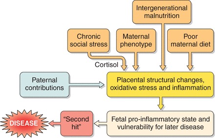
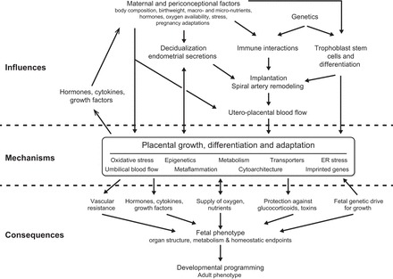
Epidemiological evidence links an individual's susceptibility to chronic disease in adult life to events during their intrauterine phase of development. Biologically this should not be unexpected, for organ systems are at their most plastic when progenitor cells are proliferating and differentiating. Influences operating at this time can permanently affect their structure and functional capacity, and the activity of enzyme systems and endocrine axes. It is now appreciated that such effects lay the foundations for a diverse array of diseases that become manifest many years later, often in response to secondary environmental stressors. Fetal development is underpinned by the placenta, the organ that forms the interface between the fetus and its mother. All nutrients and oxygen reaching the fetus must pass through this organ. The placenta also has major endocrine functions, orchestrating maternal adaptations to pregnancy and mobilizing resources for fetal use. In addition, it acts as a selective barrier, creating a protective milieu by minimizing exposure of the fetus to maternal hormones, such as glucocorticoids, xenobiotics, pathogens, and parasites. The placenta shows a remarkable capacity to adapt to adverse environmental cues and lessen their impact on the fetus. However, if placental function is impaired, or its capacity to adapt is exceeded, then fetal development may be compromised. Here, we explore the complex relationships between the placental phenotype and developmental programming of chronic disease in the offspring. Ensuring optimal placentation offers a new approach to the prevention of disorders such as cardiovascular disease, diabetes, and obesity, which are reaching epidemic proportions.


Figures

FIGURE 1.
FIGURE 1.
Diagrammatic illustration showing how the placenta may modulate and transduce environmental cues that lead to developmental programming of the fetus. The functional capacity of the placenta will depend on its development and its ability to adapt, as well as any reserve that exists.
FIGURE 2.
FIGURE 2.
Diagrammatic representation of the three main processes by which materials can cross the interhemal placental membrane: diffusion, transporter-mediated, and endocytosis. The nature of the mechanism involved will determine how readily the placenta can adapt to facilitate transport under adverse conditions.
FIGURE 3.
FIGURE 3.
Diagrammatic representation of the gross morphology of the placenta and the histology of the interhemal membrane in the human, mouse, and sheep. In each case, the bottom panel represents detail of the area outlined by the square in the top panel. A: in the human, the fetal villi arise as a series of lobules (L) from the chorionic plate (CP). The basal plate abutting the maternal decidua (D) is thrown into a series of folds forming septae (S) that partially compartmentalize the placenta into lobes. Each lobe may contain one or more lobules. Maternal blood enters the intervillous space (IVS) from the spiral arteries (SA), passes between the villi, and drains into the openings of the uterine veins on the septae. B: a single layer of syncytiotrophoblast (Stb) covers each villus and is generated from underlying cytotrophoblast (Ctb) cells. It is bathed by maternal blood in the IVS from the start of the 2nd trimester onwards. Fetal capillaries (FC) within the stromal core (Str) invaginate to reduce the length of the diffusion pathway (arrowed). C: the mouse placenta is divided into an exchange labyrinth zone (Lz) and an endocrine junctional zone (Jz). The visceral endoderm layer of the inverted yolk sac (YS) is exposed to the decidua (D) after the outer parietal layer breaks down (dotted line). This represents an important route of nutrient exchange during early pregnancy and may continue until term. D: in the labyrinth, the syncytiotrophoblast (Stb) is two-layered, and an additional layer of sinusoidal giant cells (SGC) lines the maternal blood spaces (MBS). Little stromal tissue (Str) is interposed between the fetal capillaries (FC) and the trophoblast. E: in sheep, fetal villi (FV) interdigitate with maternal crypts within specialized areas of the endometrium (E), the caruncles, to form placentomes. In between placentomes, the trophoblast forms areolae (Ar) opposite the openings of the endometrial glands (EG). Histotroph from the glands is taken up by the trophoblast, representing another route for maternal-fetal transfer. F: within a placentome, there are six tissue layers interposed between the maternal (MC) and fetal (FC) capillaries: the maternal endothelium, maternal stromal tissue (MStr), the uterine epithelium which is converted into a synepithelium by the migration and fusion of fetal binucleate cells, the trophoblast (Tr), the fetal stroma (FStr), and the fetal endothelial cells. Differences in the nature of the interhemal interface mean that extrapolation of transport data from one species to another may not always be justified.
FIGURE 4.
FIGURE 4.
The relationship between the vascular plexuses of the secondary yolk sac and the chorioallantoic placenta, and the developing heart. Because these two beds account for a substantial portion of the total vascular impedance to flow sensed by the embryonic heart, poor vascularity in these organs would offer an increased load to the heart, altering gene expression patterns and leading to congenital defects or a myocardium that is vulnerable for later disease. (Image is reprinted from www.netterimages.com, with permission.)
FIGURE 5.
FIGURE 5.
Coronary heart disease mortality in 2,571 men born in Sheffield, UK, during 1907–1930 as a function of the placental-to-birth weight ratio expressed as a percentage. The lowest rates of death from heart disease were found among men where the placental weight was ∼19% of the newborn body weight (P = 0.03). [Adapted from Godfrey (217), with permission from Elsevier.]
FIGURE 6.
FIGURE 6.
Birth and placental weights of 17,000 live births in Unizah, Saudi Arabia. The points in the top left box represent relatively low placental weights associated with relatively large babies, which have been defined as efficient placentas. The bottom right box shows low efficiency placentas where large placentas nourished low-birth-weight babies. These two extremes of efficiency may represent different kinds of programming. [Adapted from Alwasel et al. (11), with permission from Elsevier.]
FIGURE 7.
FIGURE 7.
Schematic representation of how multiple environments may give rise to placental metaflammation or “cold, smoldering inflammation,” and how this may predispose the fetus to chronic disease.
FIGURE 8.
FIGURE 8.
In the Helsinki Birth Cohort, hypertension is related to the surface area of the delivered placenta, in mothers of below median height (160 cm) (P = 0.002) but not for tall mothers (P = 0.72). [From Thornburg et al. (531), with permission from Elsevier, using data from Barker et al. (39).]
FIGURE 9.
FIGURE 9.
A summary of the principal mechanisms for oxygen sensing in cells and of the effects of modulating oxygen concentration on cell behavior that have been reported for the placenta.
FIGURE 10.
FIGURE 10.
Diagrammatic summary of the principal ways by which the Unfolded Protein Response pathway may interact with the mTOR/AKT pathway to modulate protein synthesis within the placenta. Both pathways receive input at various levels regarding oxygen and nutrient availability and will influence cell proliferation and growth. See text for details.
FIGURE 11.
FIGURE 11.
Schematic summary showing how various environmental influences may interact with, and be modulated by, the placenta and the consequences for developmental programming of the fetus.

References

    1. Acosta O, Ramirez VI, Lager S, Gaccioli F, Dudley DJ, Powell TL, Jansson T. Increased glucose and placental GLUT-1 in large infants of obese nondiabetic mothers. Am J Obstet Gynecol 212: e221–227, 2015. - PubMed
    1. Acosta-Alvear D, Zhou Y, Blais A, Tsikitis M, Lents NH, Arias C, Lennon CJ, Kluger Y, Dynlacht BD. XBP1 controls diverse cell type- and condition-specific transcriptional regulatory networks. Mol Cell 27: 53–66, 2007. - PubMed
    1. Adams Waldorf KM, McAdams RM. Influence of infection during pregnancy on fetal development. Reproduction 146: R151–162, 2013. - PMC - PubMed
    1. Adamson SL, Lu Y, Whiteley KJ, Holmyard D, Hemberger M, Pfarrer C, Cross JC. Interactions between trophoblast cells and the maternal and fetal circulation in the mouse placenta. Dev Biol 250: 358–373, 2002. - PubMed
    1. Alastalo H, Raikkonen K, Pesonen AK, Osmond C, Barker DJ, Kajantie E, Heinonen K, Forsen TJ, Eriksson JG. Cardiovascular health of Finnish war evacuees 60 years later. Ann Med 41: 66–72, 2009. - PubMed

Publication types

LinkOut - more resources