

Expressions of Collagen I and III in Hypoxic Keloid Tissue
- PMID: 27604536
- PMCID: PMC5425142
Expressions of Collagen I and III in Hypoxic Keloid Tissue
Abstract
Background: Wound heals itself spontaneously as physiological process. However, in some individuals, small wounds such as parenteral injections or body piercings may cause increased expression of collagen synthesis. The condition is known as keloid. Histopathology of keloid demonstrates extensive tissue proliferation that extends beyond the margin of primary wound. As a result, it develops uncontrolled or excessive fibrogenesis and tremendous source of collagen that still causes clinical problems until now. A wound, no matter how small the size is, will be followed by increased expression of collagen synthesis. Procollagen I and III is one of markers indicating the development of fibrosis. In fibrosis, there is hypoxia, which is characterized by stabilization of HIF-1α. Therefore, our study was aimed to obtain information about expression of collagen I and III in hypoxic keloid tissue.
Method: The study design was observational descriptive. Keloid specimens were obtained from biopsy and preputium skins as the control specimens were obtained from circumcision. There were 10 tissue specimens for each specimen group. The analysis performed were evaluation of mRNA expression on collagen I, collagen III and HIF-1α using RT-PCR, the evaluation of HIF-1α protein level using ELISA and the expression of collagen I and collagen III protein using immunohistochemistry. Statistically, data was analyzed by unpaired t-test.
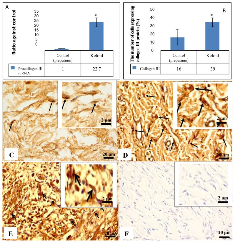
Results: In keloid with excessive cell proliferation, we found that the expression of procollagen I mRNA increased 35 times and the expression of procollagen III mRNA increased 27.1 times compared to preputium control group (p<0.05). The expression of procollagen I protein in the dermal layer of keloid was 61% and in the preputium was 37% (p<0.05). The expression of collagen III protein in the dermal layer of keloid was 39% and in the preputium was 16% (p<0.05). There was a 5-fold increase on expression of HIF-1α mRNA in keloid tissue compared to those in preputium (p<0.05). The levels of HIF-1α protein in keloid tissue was 0.201 ng/mg protein and the level in preputium was 0.122 ng/mg protein (p<0.05). There was a strong positive and extremely significant correlation between the expression of HIF-1α protein and procollagen III (R=0.744; p<0.05, Pearson), but HIF-1α with procollagen I are weak correlation (R=0.360; p>0.05, Pearson) Conclusion: Expression of collagen I and III have important role in hypoxic keloid tissue characterized by increased expressions. The expression of collagen I and III is associated with stable HIF-1α in keloid tissue.
Keywords: HIF-1α; Hypoxia; collagen; keloid.
Figures


Similar articles
-
Increased periostin expression affects the proliferation, collagen synthesis, migration and invasion of keloid fibroblasts under hypoxic conditions.Int J Mol Med. 2014 Jul;34(1):253-61. doi: 10.3892/ijmm.2014.1760. Epub 2014 Apr 25. Int J Mol Med. 2014. PMID: 24788198
-
Role of Hypoxia Inducible Factor-1 Alpha (HIF-1α) in Cytoglobin Expression and Fibroblast Proliferation of Keloids.Kobe J Med Sci. 2019 May 22;65(1):E10-E18. Kobe J Med Sci. 2019. PMID: 31341152 Free PMC article.
-
Keloid-derived keratinocytes acquire a fibroblast-like appearance and an enhanced invasive capacity in a hypoxic microenvironment in vitro.Int J Mol Med. 2015 May;35(5):1246-56. doi: 10.3892/ijmm.2015.2135. Epub 2015 Mar 13. Int J Mol Med. 2015. PMID: 25777304 Free PMC article.
-
Role of HIF-1α in pathogenic mechanisms of keloids.J Cosmet Dermatol. 2023 May;22(5):1436-1448. doi: 10.1111/jocd.15601. Epub 2023 Jan 31. J Cosmet Dermatol. 2023. PMID: 36718786 Review.
-
Hypoxia-inducible factor 1-alpha (HIF-1α) as a factor mediating the relationship between obesity and heart failure with preserved ejection fraction.Obes Rev. 2019 May;20(5):701-712. doi: 10.1111/obr.12828. Epub 2019 Mar 3. Obes Rev. 2019. PMID: 30828970 Review.
Cited by
-
Is the Mitochondrial Function of Keloid Fibroblasts Affected by Cytoglobin?Malays J Med Sci. 2021 Apr;28(2):39-47. doi: 10.21315/mjms2021.28.2.4. Epub 2021 Apr 21. Malays J Med Sci. 2021. PMID: 33958959 Free PMC article.
-
CircRNA_0263 and circRNA_1507 are dysregulated in a rat model of atrial fibrosis induced by chronic intermittent hypoxia.Am J Transl Res. 2023 Jan 15;15(1):63-81. eCollection 2023. Am J Transl Res. 2023. PMID: 36777857 Free PMC article.
-
The deubiquitinating enzyme USP37 promotes keloid fibroblasts proliferation and collagen production by regulating the c-Myc expression.Int Wound J. 2023 May;20(5):1517-1524. doi: 10.1111/iwj.14006. Epub 2022 Nov 4. Int Wound J. 2023. PMID: 36333840 Free PMC article.
-
Roles of the HIF-1α pathway in the development and progression of keloids.Heliyon. 2023 Jul 25;9(8):e18651. doi: 10.1016/j.heliyon.2023.e18651. eCollection 2023 Aug. Heliyon. 2023. PMID: 37636362 Free PMC article. Review.
-
CD44-dependent inflammation, fibrogenesis, and collagenolysis regulates extracellular matrix remodeling and tensile strength during cutaneous wound healing.Matrix Biol. 2019 Jan;75-76:314-330. doi: 10.1016/j.matbio.2018.06.004. Epub 2018 Jun 9. Matrix Biol. 2019. PMID: 29894820 Free PMC article. Review.
References
-
- Morris PJ, Wood WC. Text Book of Surgery. 2nd Ed. Oxford University Press; New York: 2000.
-
- Saed GM, Ladin D, Olson J, Han X, Hou Z, Fivenson D. Analysis of p53 Gene Mutations in Keloids Using Polymerase Chain Reaction–Based Single-Strand Conformational Polymorphism and DNA Sequencing. Arch dermatol. 1998;134:963–71. - PubMed
-
- Lei ZX, Yin JD, Chang WJ, Li LJ, Zhong LZ, Long CJ. Transforming growth factor-α1 phage model peptides isolated from a phage display 7-mer peptide library can inhibit the activity of keloid fibroblasts. Chin Med J. 2011;124(3):429–435. - PubMed
Publication types
MeSH terms
Substances
LinkOut - more resources
Full Text Sources
Research Materials