. 2016 Sep;28(9):2097-2116.
doi: 10.1105/tpc.16.00490. Epub 2016 Sep 7.

MYB107 and MYB9 Homologs Regulate Suberin Deposition in Angiosperms

Affiliations

MYB107 and MYB9 Homologs Regulate Suberin Deposition in Angiosperms

Justin Lashbrooke et al. Plant Cell. 2016 Sep.

Abstract

Suberin, a polymer composed of both aliphatic and aromatic domains, is deposited as a rough matrix upon plant surface damage and during normal growth in the root endodermis, bark, specialized organs (e.g., potato [Solanum tuberosum] tubers), and seed coats. To identify genes associated with the developmental control of suberin deposition, we investigated the chemical composition and transcriptomes of suberized tomato (Solanum lycopersicum) and russet apple (Malus x domestica) fruit surfaces. Consequently, a gene expression signature for suberin polymer assembly was revealed that is highly conserved in angiosperms. Seed permeability assays of knockout mutants corresponding to signature genes revealed regulatory proteins (i.e., AtMYB9 and AtMYB107) required for suberin assembly in the Arabidopsis thaliana seed coat. Seeds of myb107 and myb9 Arabidopsis mutants displayed a significant reduction in suberin monomers and altered levels of other seed coat-associated metabolites. They also exhibited increased permeability, and lower germination capacities under osmotic and salt stress. AtMYB9 and AtMYB107 appear to synchronize the transcriptional induction of aliphatic and aromatic monomer biosynthesis and transport and suberin polymerization in the seed outer integument layer. Collectively, our findings establish a regulatory system controlling developmentally deposited suberin, which likely differs from the one of stress-induced polymer assembly recognized to date.


Figures

Figure 1.
Figure 1.
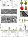
Silencing of SlDCR in Tomato Leads to Suberization of the Fruit Surface. (A) Examination of SlDCR expression in developing tomato fruit via RT-qPCR shows a skin enriched expression profile. IG, immature green; MG, mature green; Br, breaker; Or, orange and red fruit stages. Error bars show se (n = 3; **P value < 0.01, Student’s t test). (B) Confirmation of SlDCR silencing in mature green stage skin tissue via RT-qPCR of in transgenic lines (SlDCR-RNAi). Error bars show se (n = 3; **P value < 0.01, Student’s t test). (C) Fruit surface phenotype of SlDCR-RNAi fruit at the mature green and red developmental stage. The disrupted epidermal layer of the SlDCR-RNAi fruit is revealed by scanning electron microscopy (see inset). (D) Scanning electron microscopy analysis of red stage fruit surface shows microcracking between SlDCR-RNAi epidermal cells. (E) Transmission electron microscopy analysis of fruit epidermal cell at the red development stage shows reduced cuticle layer (cl) and an increase in lipid inclusion bodies (li) in the cytosol of SlDCR-RNAi cells. (F) Chemical analysis via gas chromatography-mass spectrometry (GC-MS) of the fruit surface polymer in SlDCR-RNAi lines shows a massive decrease in C16-9/10,16-DHFA and an increase in monomers typically found in suberin polymer. Error bars show se (n = 4; **P value < 0.01, Student’s t test). (G) Expression analysis (RT-qPCR) shows a massive increase in expression of SlASFT and SlGPAT5 (over 350-fold) in SlDCR-RNAi mature green fruit skin. Error bars show se (n = 3; **P value < 0.01, Student’s t test).
Figure 2.
Figure 2.
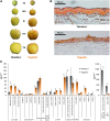
Surface Phenotype and Chemical Composition of Apple Russet. (A) Two Golden Delicious apple clones were analyzed: the normal skinned ‘Reinders’ clone and the russeted ‘Rugiada’ clone. ‘Rugiada’ displays a rough, brown, and cracked fruit surface. EG, early green; MG, mature green; Br, breaker; Har, harvest; PH, postharvest fruit stages. (B) Light microscopy of the epidermal layer of harvest stage fruit from ‘Reinders’ and ‘Rugiada’ using the lipid stain Sudan IV shows a dramatic reduction in cuticle deposition in the latter clone. (C) Chemical analysis via GC-MS of the surface polymer of these two clones highlights the dramatic reduction in C16-9/10,16-DHFA and C16-ω-HFA monomers of ‘Rugiada’, while an increase in longer chain length C20- and C22-ω-HFA is observed, together with an increase in aromatics. Error bars show se (n = 4; *P value < 0.05, **P value < 0.01, Student’s t test).
Figure 3.
Figure 3.
Orthologous Genes Upregulated in Both the Suberized Fruit Skin Tissues of SlDCR-RNAi Tomato and Russeted Apple. Genes upregulated in SlDCR-RNAi compared with the wild type and in the russeted ‘Rugiada’ clone compared with the regular skinned ‘Reinders’ were depicted in a heat map. For a full list of identified genes and the corresponding expression patterns, see Supplemental Data Sets 2 and 3 and Supplemental Table 2. Bar indicates log2(fold change) of gene expression centered on the mean of expression for each gene. EG, early green; MG, mature green; Br, breaker; Har, harvest; PH, postharvest fruit stages.
Figure 4.
Figure 4.
A Multispecies Gene Expression Signature for Suberin Biosynthesis. Orthologous genes identified through the multi-species suberin gene coexpression analysis are represented graphically. Genes found to be coexpressed with baits representing known suberin synthesis genes were identified in seven data sets and compared for orthology. Clockwise from top right the images represent transcriptomics data sets that examined expression in SlDCR-RNAi versus wild-type fruit skin tissues, russeted versus regular skinned apples surface tissues, multiple tissue types from whole tomato plant, Arabidopsis seeds tissues, waterlogged rice roots versus wild-type roots, whole grapevine plant tissues, and whole potato plant tissues. Edges (blue lines) represent presence of the corresponding ortholog to the expression data set, while nodes (red circles) represent orthologous gene groups. Bar indicates the number of experiments in which the orthologous gene group was identified (i.e., four, five, six, or seven coexpression experiments; seven out of seven analyses is represented by the largest and most intensely red colored circle). Orthologs were grouped based on their proposed function. For respective gene IDs and details of the coexpression parameters, see Table 1 and Supplemental Table 6, respectively.
Figure 5.
Figure 5.
Identification of a Clade of MYB Factors Likely Involved in Suberin Formation across Multiple Species. Molecular phylogenetic analysis of Arabidopsis MYB factors from subgroups 9, 10, 11, and 24 was performed together with the MYB factors identified in this study to be expressed in suberin forming tissues of other species (in bold). While subgroups 9 and 11 form separate clades, division of subgroups 10 and 24 was not clearly resolved. Protein domain analysis identified three C-terminal motifs specific to this combined clade that may be involved in regulation of suberin formation (SUB-I, SUB-II, and SUB-III). The motifs are marked on top of the gene structure downstream of the indicated R2-R3 domains characteristic of plant MYB proteins. Detailed representation of the identified motifs is shown in full below. AtMYB9 and AtMYB107 are highlighted in red and bold text. ClustalW and MEGA6 software were used to align the proteins and compute the neighbor-joining tree with significance percentages (bootstrap values out of 1000). The alignment used to generate the phylogeny is presented in Supplemental Data Set 6.
Figure 6.
Figure 6.
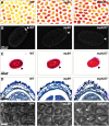
Seed Coat Phenotypes of myb107 and myb9 Arabidopsis Mutant Lines. (A) Tetrazolium salt penetration assays for seeds of Arabidopsis T-DNA insertion lines identified an increase in seed permeability (resulting in red staining of seeds) for myb107 and myb9 mutant lines when compared with the Col-0 wild-type control. (B) Autofluorescence of suberin in mature Arabidopsis seeds when excited at 365 nm shows a decrease in fluorescence in the hilum region (arrowheads) of the mutant seeds. (C) Ruthenium red staining of seed mucilage highlights the decreased mucilage layer (arrowheads) of myb107 and myb9 seeds. (D) Histological sections of seed coats stained with toluidine blue O shows the relative disorganization of the integument layers in the myb107 seed coat. (E) Scanning electron microscopy images of seed coat surface structure and columella cells identifies gaps at the columella cell borders of myb107 seeds (arrowheads).
Figure 7.
Figure 7.
Chemical Analysis of Seed Coat Polymer from myb9 and myb107 Mutant Lines. Chemical analysis via GC-MS of the depolymerized seed coat polymer was performed for mature dry seeds from myb9 and myb107 Arabidopsis T-DNA insertion lines. A decrease in aliphatic and aromatic constituents when compared with Col-0 wild type can be observed. Error bars represent se (n = 3; *P value < 0.05, **P value < 0.01, Student’s t test).
Figure 8.
Figure 8.
Decreased Germination Rates Were Observed for myb107 Seeds under Osmotic and Salt Stress. Germination rates of wild-type (Col-0), myb9, and myb107 mature seeds on control conditions ([A]; 0.5% Murashige and Skoog medium/Suc), mild osmotic stress ([B];150 mM mannitol), severe osmotic stress ([C]; 300 mM mannitol), mild salt stress ([D]; 100 mM NaCl), and severe salt stress ([E]; 200 mM NaCl) are shown. Horizontal axis represents days after imbibition. See Supplemental Figure 17 for photographs representing characteristic germination phenotypes under these conditions 8 d after imbibition. Successful germination was scored according to radical emergence (as illustrated in Supplemental Figure 17). Error bars show se (n = 5, 20 seeds per replicate; *P value < 0.05, Student’s t test).
Figure 9.
Figure 9.
Model for Suberin Formation in the Arabidopsis Seed Coat and the Role of MYB9 and MYB107 Transcription Factors. MYB9 and MYB107 (represented in bold text) appear to act in tandem to positively regulate various aspects of suberin formation (labeled in bold italics) in the outer integument layer of the seed coat, including fatty acid and phenylpropanoid biosynthesis, and extracellular suberin monomer transport and polymerization. A feedback loop likely exists between MYB9 and MYB107. Proposed downstream targets of MYB107 and MYB9 are shown with blue and red arrows, respectively. The hypothesis is supported by data from the chemical analysis of the seed coat polyester, gene expression data from seeds, as well as seed permeability assays. FA, fatty acid; VLCFAs, very-long-chain fatty acids; GPAT5, GLYCEROL-3-PHOSPHATE ACYLTRANSFERASE5; ASFT, ALIPHATIC SUBERIN FERULOYL TRANSFERASE; KCS17, 3-KETOACYL-COA SYNTHASE17; 4CL5, 4-COUMARATE:COA LIGASE5; DFR-like1, DIHYDROFLAVONOL 4-REDUCTASE-LIKE1; LTPG5, GLYCOSYLPHOSPHATIDYLINOSITOL-ANCHORED LIPID PROTEIN TRANSFER5; SUS, SUBERIN SYNTHASE.

References

    1. Alonso J.M., et al. (2003). Genome-wide insertional mutagenesis of Arabidopsis thaliana. Science 301: 653–657. - PubMed
    1. Bailey T.L., Boden M., Buske F.A., Frith M., Grant C.E., Clementi L., Ren J., Li W.W., Noble W.S. (2009). MEME SUITE: tools for motif discovery and searching. Nucleic Acids Res. 37: W202–W208. - PMC - PubMed
    1. Bargel H., Koch K., Cerman Z., Neinhuis C. (2006). Structure–function relationships of the plant cuticle and cuticular waxes–a smart material? Funct. Plant Biol. 33: 893–910. - PubMed
    1. Beisson F., Li-Beisson Y., Pollard M. (2012). Solving the puzzles of cutin and suberin polymer biosynthesis. Curr. Opin. Plant Biol. 15: 329–337. - PubMed
    1. Beisson F., Li Y., Bonaventure G., Pollard M., Ohlrogge J.B. (2007). The acyltransferase GPAT5 is required for the synthesis of suberin in seed coat and root of Arabidopsis. Plant Cell 19: 351–368. - PMC - PubMed