. 2016 Sep;28(9):2147-2160.
doi: 10.1105/tpc.16.00428. Epub 2016 Sep 7.

Arabidopsis STAY-GREEN, Mendel's Green Cotyledon Gene, Encodes Magnesium-Dechelatase

Affiliations

Arabidopsis STAY-GREEN, Mendel's Green Cotyledon Gene, Encodes Magnesium-Dechelatase

Yousuke Shimoda et al. Plant Cell. 2016 Sep.

Abstract

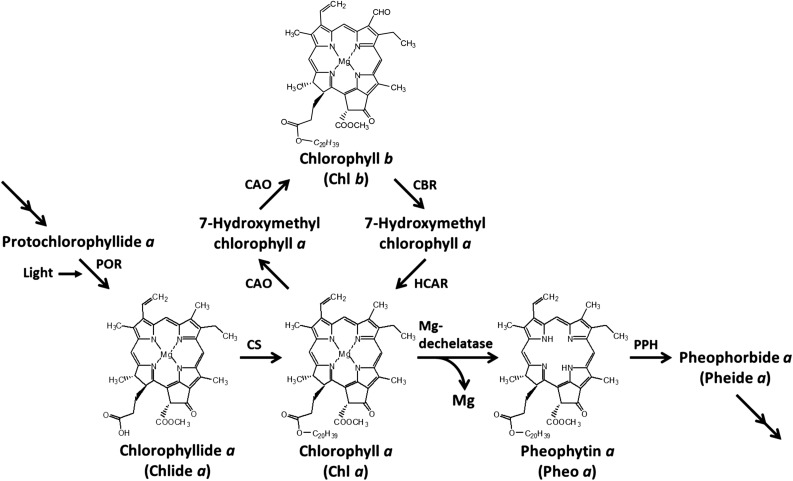
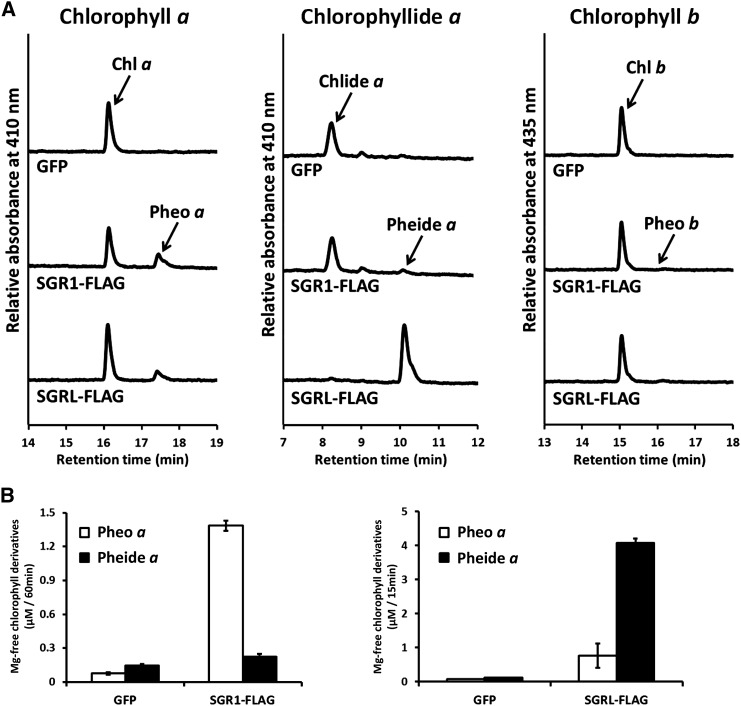
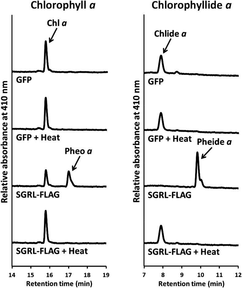
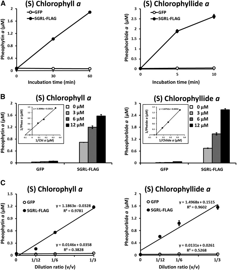
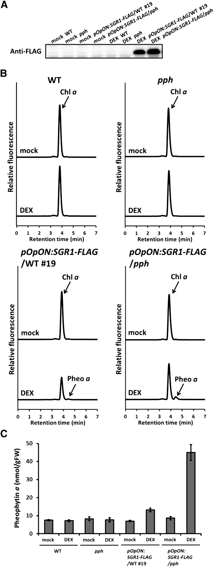
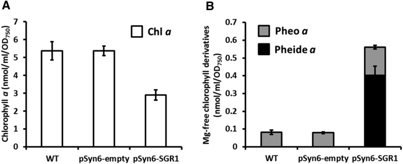
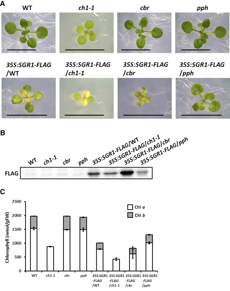
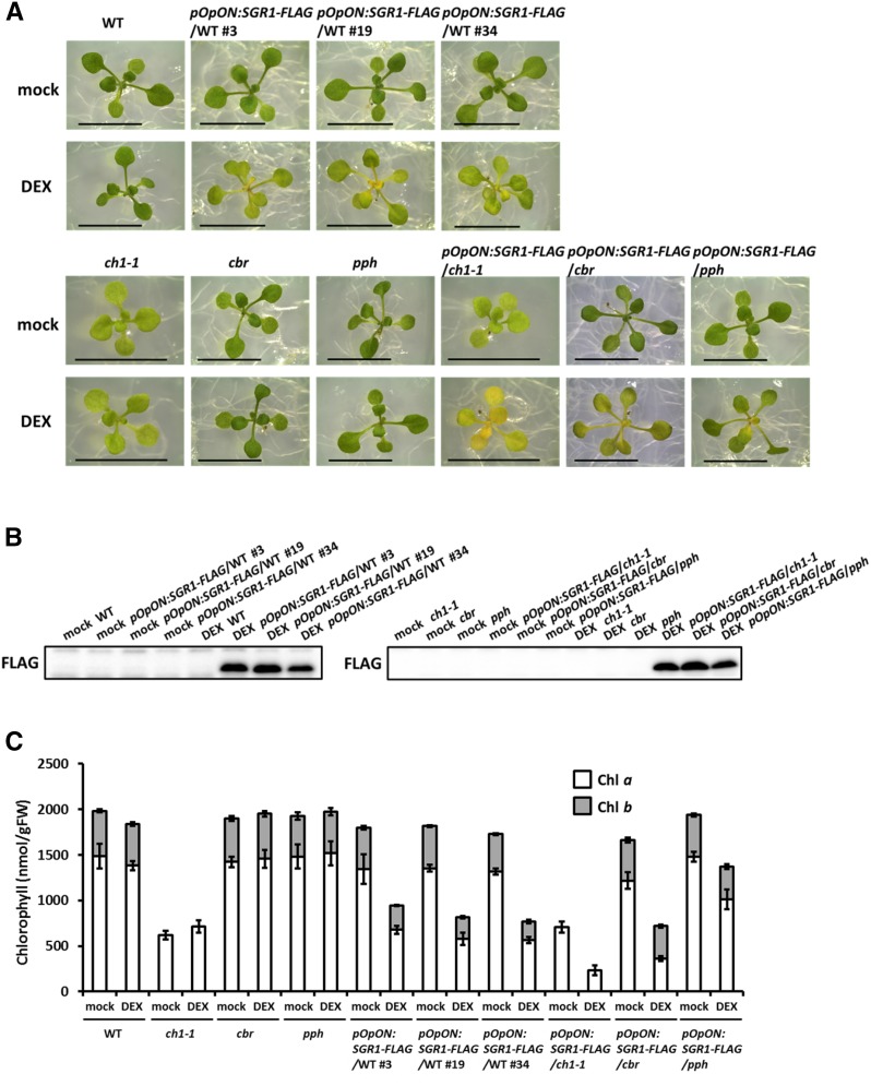
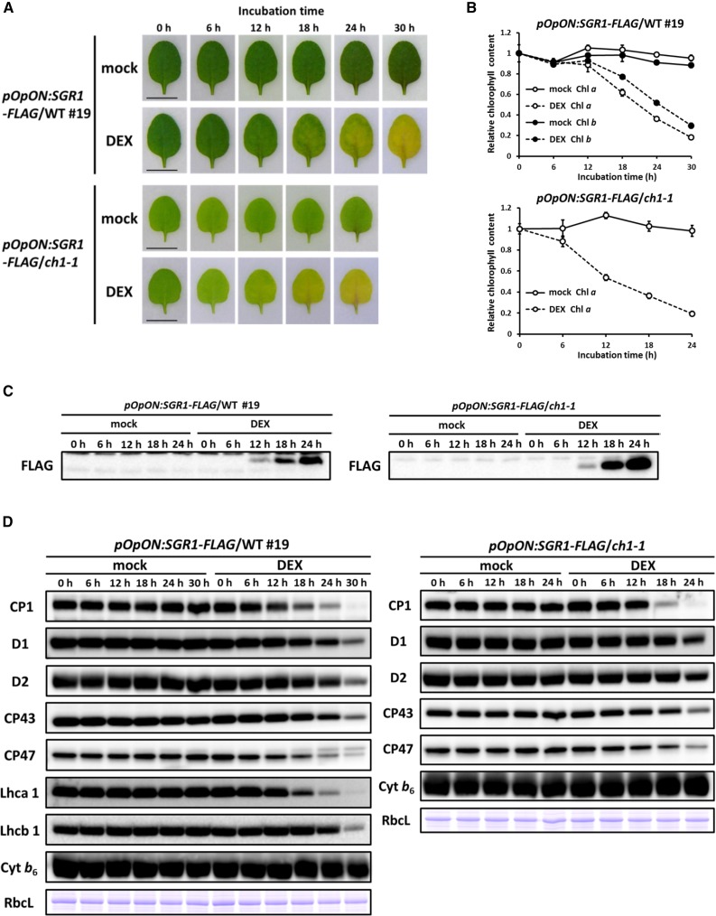
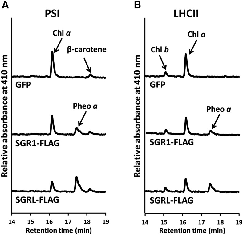
Pheophytin a is an essential component of oxygenic photosynthetic organisms because the primary charge separation between chlorophyll a and pheophytin a is the first step in the conversion of light energy. In addition, conversion of chlorophyll a to pheophytin a is the first step of chlorophyll degradation. Pheophytin is synthesized by extracting magnesium (Mg) from chlorophyll; the enzyme Mg-dechelatase catalyzes this reaction. In this study, we report that Mendel's green cotyledon gene, STAY-GREEN (SGR), encodes Mg-dechelatase. The Arabidopsis thaliana genome has three SGR genes, SGR1, SGR2, and STAY-GREEN LIKE (SGRL). Recombinant SGR1/2 extracted Mg from chlorophyll a but had very low or no activity against chlorophyllide a; by contrast, SGRL had higher dechelating activity against chlorophyllide a compared with chlorophyll a All SGRs could not extract Mg from chlorophyll b Enzymatic experiments using the photosystem and light-harvesting complexes showed that SGR extracts Mg not only from free chlorophyll but also from chlorophyll in the chlorophyll-protein complexes. Furthermore, most of the chlorophyll and chlorophyll binding proteins disappeared when SGR was transiently expressed by a chemical induction system. Thus, SGR is not only involved in chlorophyll degradation but also contributes to photosystem degradation.


Figures

Figure 1.
Figure 1.
Chlorophyll Metabolic Pathway in Land Plants. Mg-dechelatase was identified in this study. CAO, chlorophyllide a oxygenase; CBR, chlorophyll b reductase; CS, chlorophyll synthase; HCAR, 7-hydroxymethyl chlorophyll a reductase; PPH, pheophytin pheophorbide hydrolase; POR, NADPH:protochlorophyllide oxidoreductase.
Figure 2.
Figure 2.
Mg-Dechelating Activity and Substrate Specificity of Recombinant SGR1 and SGRL. (A) Pigment analysis after incubation of chlorophyll derivatives with SGR. Chlorophyll a and chlorophyllide a were incubated with recombinant GFP, SGR1 with a FLAG-tag (SGR1-FLAG) for 60 min, or with recombinant SGRL with a FLAG-tag (SGRL-FLAG) for 15 min. Recombinant proteins were prepared with a wheat germ protein expression system and diluted 3-fold with the reaction buffer without purification. GFP was used as a negative control because it has a similar molecular weight as SGR. Chlorophyll b was incubated with recombinant GFP, SGR1-FLAG, and SGRL-FLAG for 60 min. The concentration of substrates was 6 µM. After incubation, pigments were analyzed using HPLC. Pigments were detected at 410 nm for chlorophyll a derivatives or 435 nm for chlorophyll b derivatives. (B) An increase in chlorophyll derivatives by SGR activity. The levels of pheophytin a and pheophorbide a were determined after incubation of recombinant GFP and SGR1 with a FLAG-tag (SGR1-FLAG and SGRL-FLAG) with 6 µM of chlorophyll a and chlorophyllide a (n = 3 ±sd). The incubation times of SGR1-FLAG and SGRL-FLAG were 60 and 15 min, respectively. Recombinant proteins were prepared with a wheat germ protein expression system and diluted with the reaction buffer without purification. GFP was used as a negative control because it has similar molecular weight as SGR.
Figure 3.
Figure 3.
Mg-Dechelating Activity of Heat-Denatured SGRL. Recombinant GFP and SGRL-FLAG were denatured by heat treatment for 5 min at 95°C. Chlorophyll a and chlorophyllide a were incubated with nondenatured or denatured recombinant GFP and SGRL-FLAG for 60 min at 25°C. Recombinant proteins were prepared by a wheat germ protein expression system and diluted 3-fold with the reaction buffer without purification. GFP was used as a negative control because it has similar molecular weight as SGR. The concentration of substrates was 6 µM. After incubation, pigments were analyzed using HPLC. Pigments were detected at 410 nm.
Figure 4.
Figure 4.
Biochemical Analysis of SGRL. (A) Time-dependent formation of Mg-free chlorophyll derivatives by SGRL-FLAG. Chlorophyll a or chlorophyllide a were incubated with recombinant GFP (open circles) and SGRL-FLAG (closed circles) for up to 60 min or 10 min at 25°C, respectively. Recombinant proteins were prepared by a wheat germ protein expression system and diluted 3-fold with the reaction buffer without purification. GFP was used as a negative control because it has similar molecular weight as SGR. The concentration of substrates was 6 µM. After incubation, the level of pheophytin a and pheophorbide a was determined using HPLC (n = 3 ±sd). (B) Kinetic analysis of Mg-dechelating of SGRL-FLAG. Various concentrations of chlorophyll a or chlorophyllide a were incubated with recombinant GFP and SGRL-FLAG for 30 or 5 min at 25°C, respectively. Recombinant proteins were prepared by a wheat germ protein expression system and diluted 3-fold with the reaction buffer without purification. GFP was used as a negative control because it has similar molecular weight as SGR. After incubation, the level of pheophytin a and pheophorbide a were determined using HPLC (n = 3 ±sd). The inset shows Lineweaver-Burk plot of kinetic data of Mg-dechelating of SGRL-FLAG. (C) SGRL-FLAG concentration-dependent formation of Mg-free chlorophyll derivatives. Chlorophyll a or chlorophyllide a were incubated with various concentrations of recombinant GFP (open circles) and SGRL-FLAG (closed circles) for 30 or 5 min at 25°C, respectively. Translation solutions containing expressed GFP and SGRL-FLAG were diluted 3, 6, or 12 times in 50 μL of reaction buffer. GFP was used as a negative control because it has similar molecular weight as SGR. The concentration of substrates was 6 µM. After incubation, the levels of pheophytin a and pheophorbide a were determined using HPLC (n = 3 ±sd).
Figure 5.
Figure 5.
SGR1 Functions as a Mg-Dechelatase in Cells. (A) SGR1 accumulation in the transformants. Inducible SGR1 with a FLAG-tag (SGR1-FLAG) was introduced into wild-type (pOpON:SGR1-FLAG/WT #19) and pph (pOpON:SGR1-FLAG/pph) plants. SGR1- FLAG was induced by DEX application in the transformants. After DEX or mock treatment for 24 h, proteins were extracted from the plants and SGR1 was detected by immunoblotting analysis using an anti-FLAG antibody. (B) Pigment analysis after SGR1 induction. After DEX or mock treatment for 24 h, pigments were extracted from the plants and analyzed using HPLC. Fluorescence intensity was monitored (410 nm excitation; 680 nm fluorescence). (C) Pheophytin a contents in the transformants. After DEX or mock treatment for 24 h, pigments were extracted from the plants and the amount of pheophytin a was determined (n = 4 ±sd).
Figure 6.
Figure 6.
SGR1 Functions in Synechococcus. (A) Chlorophyll a contents of Synechococcus. Chlorophyll a content of Synechococcus harboring the pSyn6 vector (pSyn6-empty) or SGR1 cloned into the pSyn6 vector (pSyn6-SGR1) was determined (n = 3 ±sd). Pigment content is shown based on OD750. (B) Derivatives of chlorophyll a in Synechococcus. Pheophytin a and pheophorbide a contents of the wild type and transformed Synechococcus were determined (n = 3 ±sd).
Figure 7.
Figure 7.
SGR1 Overexpression in Arabidopsis. (A) Visual phenotype of the transformants. SGR1 with a FLAG-tag (SGR1-FLAG) was overexpressed in wild-type plants and in the ch1-1, cbr, and pph mutants. Bars = 1 cm. (B) SGR1 accumulation in the transformants. Proteins were extracted from the plants and SGR1 was detected by immunoblotting analysis using an anti-FLAG antibody. (C) Chlorophyll content of the transformants. Chlorophyll was extracted from the plants and the amount of chlorophyll a and b was determined (n = 3 ±sd).
Figure 8.
Figure 8.
SGR1 Induction in Arabidopsis. (A) Visual phenotype of the transformants. SGR1 with a FLAG-tag (SGR1-FLAG) was induced by DEX application for 24 h in wild-type plants and in the ch1-1, cbr, and pph mutants grown for 2 weeks. Three independent transformants in a wild-type background are shown. Bars = 1 cm. (B) SGR1 accumulation in the transformants. Proteins were extracted from the plants and SGR1 was detected by immunoblotting analysis using an anti-FLAG antibody. (C) Chlorophyll contents of the transformants. Chlorophyll was extracted from the plants and the amount of chlorophyll a and b was determined (n = 4 ±sd).
Figure 9.
Figure 9.
Degradation of Chlorophyll and Chlorophyll Binding Protein by the Induction of SGR1. (A) Color changes of leaves. Inducible SGR1 with a FLAG-tag was introduced into wild-type (pOpON:SGR1-FLAG/WT #19) or ch1-1 (pOpON:SGR1-FLAG/ch1-1) plants. DEX- or mock-treated excised leaves were observed for up to 30 h. Bars = 0.5 cm. (B) Chlorophyll contents of leaves. Chlorophyll contents of pOpON:SGR1-FLAG/WT #19 and pOpON:SGR1-FLAG/ch1-1 were determined before and after DEX or mock treatment for up to 30 h (n = 4 ±sd). Comparisons were made to a 0 h control. (C) SGR1 accumulation in leaves. Proteins were extracted from leaves and SGR1 was detected using immunoblotting analysis with an anti-FLAG antibody. (D) Chloroplast protein content in leaves. Proteins were extracted from the pOpON:SGR1-FLAG/WT #19 and pOpON:SGR1-FLAG/ch1-1 excised leaves before and after DEX or mock treatment for up to 30 h. The large subunit of ribulose-1,5-bisphosphate carboxylase/oxygenase (RbcL) was detected using Coomassie blue staining.
Figure 10.
Figure 10.
Magnesium Extraction from Chlorophyll in the Chlorophyll-Protein Complex by SGR. (A) Pigment analysis after incubation of PSI with SGR. PSI was isolated from Arabidopsis and incubated with recombinant GFP, SGR1 with a FLAG-tag (SGR1-FLAG), and SGRL with a FLAG-tag (SGRL-FLAG) for 60 min. Recombinant proteins were prepared by a wheat germ protein expression system and diluted with the same volume of reaction buffer without purification. GFP was used as a negative control because it has similar molecular weight as SGR. After incubation, pigments were analyzed using HPLC. Pigments were detected at 410 nm. (B) Pigment analysis after incubation of LHCII with SGR. LHCII was isolated from Arabidopsis and incubated with recombinant GFP, SGR1-FLAG, and SGRL-FLAG for 60 min. Recombinant proteins were prepared with a wheat germ protein expression system and diluted with same volume of the reaction buffer without purification. GFP was used as a negative control because it has a similar molecular weight as SGR. After incubation, pigments were analyzed using HPLC. Pigments were detected at 410 nm.

Comment in

References

    1. Armstead I., et al. (2007). Cross-species identification of Mendel’s I locus. Science 315: 73. - PubMed
    1. Chidgey J.W., Linhartová M., Komenda J., Jackson P.J., Dickman M.J., Canniffe D.P., Koník P., Pilný J., Hunter C.N., Sobotka R. (2014). A cyanobacterial chlorophyll synthase-HliD complex associates with the Ycf39 protein and the YidC/Alb3 insertase. Plant Cell 26: 1267–1279. - PMC - PubMed
    1. Christ B., Hörtensteiner S. (2013). Mechanism and significance of chlorophyll breakdown. J. Plant Growth Regul. 33: 4–20.
    1. Costa M.L., Civello P.M., Chaves A.R., Martínez G.A. (2002). Characterization of Mg-dechelatase activity obtained from Fragaria × ananassa fruit. Plant Physiol. Biochem. 40: 111–118.
    1. Craft J., Samalova M., Baroux C., Townley H., Martinez A., Jepson I., Tsiantis M., Moore I. (2005). New pOp/LhG4 vectors for stringent glucocorticoid-dependent transgene expression in Arabidopsis. Plant J. 41: 899–918. - PubMed