. 2017 Jan;77(1):72-81.
doi: 10.1002/pros.23250. Epub 2016 Sep 7.

TGF-β Effects on Prostate Cancer Cell Migration and Invasion Require FosB

Affiliations

TGF-β Effects on Prostate Cancer Cell Migration and Invasion Require FosB

Cachétne S X Barrett et al. Prostate. 2017 Jan.

Abstract

Background: Activator Protein-1 (AP-1) family (cJun, JunB, JunD, cFos, FosB, Fra1, and Fra2) plays a central role in the transcriptional regulation of many genes that are associated with cell proliferation, differentiation, migration, metastasis, and survival. Many oncogenic signaling pathways converge at the AP-1 transcription complex. Transforming growth factor beta (TGF-β) is a multifunctional regulatory cytokine that regulates many aspects of cellular function, including cellular proliferation, differentiation, migration, apoptosis, adhesion, angiogenesis, immune surveillance, and survival.

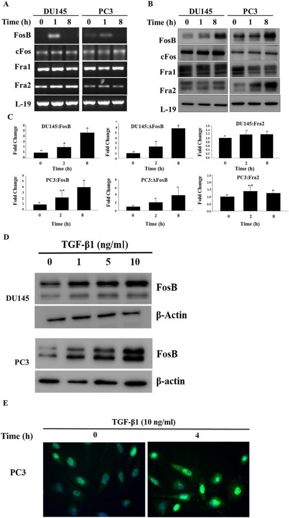
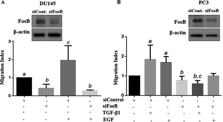
Methods: This study investigated, the role of FOS proteins in TGF-β signaling in prostate cancer cell proliferation, migration, and invasion. Steady state expression levels of FOS mRNA and proteins were determined using RT-PCR and western blotting analyses. DU145 and PC3 prostate cancer cells were exposed to TGF-β1 at varying time and dosage, RT-PCR, western blot, and immunofluorescence analyses were used to determine TGF-β1 effect on FOS mRNA and protein expression levels as well as FosB subcellular localization. Transient silencing of FosB protein was used to determine its role in cell proliferation, migration, and invasion.

Results: Our data show that FOS mRNA and proteins were differentially expressed in human prostate epithelial (RWPE-1) and prostate cancer cell lines (LNCaP, DU145, and PC3). TGF-β1 induced the expression of FosB at both the mRNA and protein levels in DU145 and PC3 cells, whereas cFos and Fra1 were unaffected. Immunofluorescence analysis showed an increase in the accumulation of FosB protein in the nucleus of PC3 cells after treatment with exogenous TGF-β1. Selective knockdown of endogenous FosB by specific siRNA did not have any effect on cell proliferation in PC3 and DU145 cells. However, basal and TGF-β1- and EGF-induced cell migration was significantly reduced in DU145 and PC3 cells lacking endogenous FosB. TGF-β1- and EGF-induced cell invasion were also significantly decreased after FosB knockdown in PC3 cells.

Conclusion: Our data suggest that FosB is required for migration and invasion in prostate cancer cells. We also conclude that TGF-β1 effect on prostate cancer cell migration and invasion may be mediated through the induction of FosB. Prostate 77:72-81, 2017. © 2016 Wiley Periodicals, Inc.

Keywords: AP-1; FosB; TGF-β; cell invasion; cell migration; prostate cancer.


Figures

Fig. 1
Fig. 1
FOS family basal expression. Steady state mRNA levels of FOS (FosB, cFos, Fra1, and Fra2) mRNA and protein expression. (A) Total RNA's were isolated and semi quantitative RT-PCR was performed to determine the mRNA levels of FosB, cFos, Fra1, and Fra2 in prostate cells. L-19 was used as an internal control. (B) Western blot analysis of FosB, cFos, Fra1, and Fra2 in prostate cells. β-actin was used as a loading control.
Fig. 2
Fig. 2
TGF-β1 effects on expression of FOS family members. (A) RT-PCR analysis of FosB, cFos, Fra1, and Fra2 mRNA levels in DU145 and PC3 prostate cancer cells after exposure to exogenous TGF-β (10 ng/ml) for different times. (B) Western blot analysis of FosB, the FosB antibody used recognizes both full length FosB (higher molecular weight band) and ΔFosB (lower molecular weight band), cFos, Fra1, and Fra2 protein levels DU145 and PC3 prostate cancer cells after exposure to exogenous TGF-β1 (10 ng/ml) for different times. (C) Band density analysis of FosB and Fra2 in DU145 and PC3 cells after treatment with TGF-β1 for 2 and 8 hr. Each band density was normalized by density of β-actin bands. Each bar represents the Mean ± SD from three independent experiments. “a and b” denote significant differences (P < 0.05) from untreated controls. (D) The Dose dependent effects of TGF-β1 on expression of FosB; Western blot analysis of FosB in prostate cancer cells DU145 and PC3 after treatment with varying concentrations of exogenous TGF-β1 (0, 1, 5, 10 ng/ml) for 4 hr. (E) Immunofluorescence, TGF-β1 activation of FosB in PC3. Cells were treated with exogenous TGF-β1 (10 ng/ml) for 0 and 4 hr.
Fig. 3
Fig. 3
Effects of FosB knock down on TGF-β1-induced cell proliferation. DU145 (A) and PC3 (B) cells were transfected with siRNA to transiently silence FosB followed by an in vitro proliferation assay.
Fig. 4
Fig. 4
Effects of FosB knock down on cell migration. Prostate cancer cells DU145 (A) and PC3 (B) were pretreated with siRNA against FosB for 72 hr. Western blots were used to confirm knock down of endogenous FosB (inserts). DU145 and PC3 cells were pretreated with siRNA against FosB, followed by treatment with 10 ng/ml of exogenous TGF-β1 and 10 ng/ml EGF migratory behavior were measured using transwell insert migration assay. Each bar represents Mean ± SEM from three independent experiments. Different letters designate statistically significant (P < 0.05) differences among different treatments.
Fig. 5
Fig. 5
Effects of FosB knock down on cell invasion. PC3 cells were pretreated with siRNA against FosB, followed by treatment with 10 ng/ml of exogenous TGF-β1 and 10 ng/ml EGF. Invasive behavior was measured using a matrigel in vitro invasion assay. Insert shows western blot used to confirm FosB knock down. Each bar represents Mean ± SEM from three independent experiments. Different letters designate statistically significant (P < 0.05) differences among different treatments.

Similar articles

Cited by

References

    1. DeSantis CE, Lin CC, Mariotto AB, Siegel RL, Stein KD, Kramer JL, Alteri R, Robbins AS, Jemal A. Cancer treatment and survivorship statistics. CA Cancer J Clin. 2014;64:252–271. - PubMed
    1. Arnold JT, Isaacs JT. Mechanisms involved in the progression of androgen-independent prostate cancers: It is not only the cancer cell's fault. Endocr Relat Cancer. 2002;9:61–73. - PMC - PubMed
    1. Collazo J, Zhu B, Larkin S, Martin SK, Pu H, Horbinski C, Koochekpour S, Kyprianou N. Cofilin drives cell-invasive and metastatic responses to TGF-beta in prostate cancer. Cancer Res. 2014;74:2362–2373. - PMC - PubMed
    1. Armstrong CM, Gao AC. Drug resistance in castration resistant prostate cancer: Resistance mechanisms and emerging treatment strategies. Am J Clin Exp Urol. 2015;3:64–76. - PMC - PubMed
    1. Chandrasekar T, Yang JC, Gao AC, Evans CP. Mechanisms of resistance in castration-resistant prostate cancer (CRPC). Transl Androl Urol. 2015;4:365–380. - PMC - PubMed

Publication types

MeSH terms

Substances