. 2017 Jan;74(2):373-392.
doi: 10.1007/s00018-016-2352-5. Epub 2016 Sep 7.

SUMOylation regulates the intracellular fate of ZO-2

Affiliations

SUMOylation regulates the intracellular fate of ZO-2

Franziska Wetzel et al. Cell Mol Life Sci. 2017 Jan.

Abstract

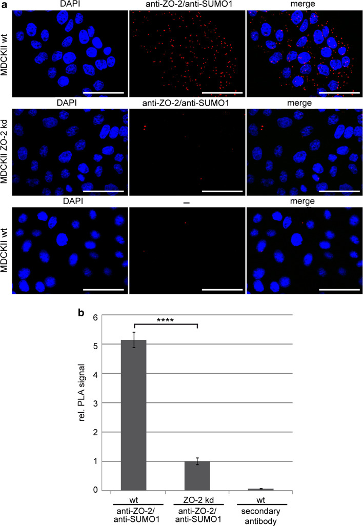
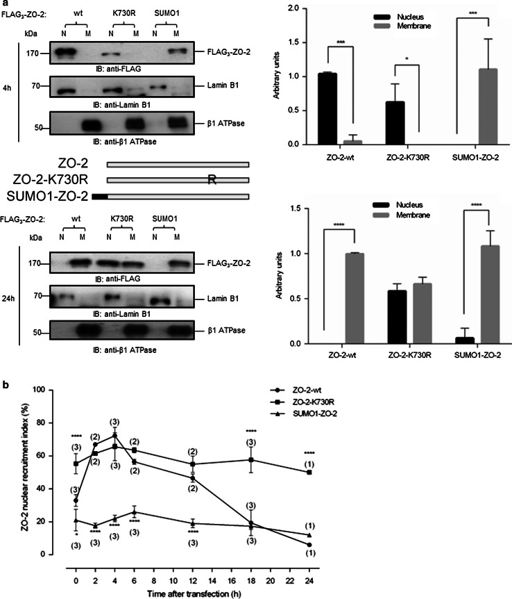
The zonula occludens (ZO)-2 protein links tight junctional transmembrane proteins to the actin cytoskeleton and associates with splicing and transcription factors in the nucleus. Multiple posttranslational modifications control the intracellular distribution of ZO-2. Here, we report that ZO-2 is a target of the SUMOylation machinery and provide evidence on how this modification may affect its cellular distribution and function. We show that ZO-2 associates with the E2 SUMO-conjugating enzyme Ubc9 and with SUMO-deconjugating proteases SENP1 and SENP3. In line with this, modification of ZO-2 by endogenous SUMO1 was detectable. Ubc9 fusion-directed SUMOylation confirmed SUMOylation of ZO-2 and was inhibited in the presence of SENP1 but not by an enzymatic-dead SENP1 protein. Moreover, lysine 730 in human ZO-2 was identified as a potential modification site. Mutation of this site to arginine resulted in prolonged nuclear localization of ZO-2 in nuclear recruitment assays. In contrast, a construct mimicking constitutive SUMOylation of ZO-2 (SUMO1ΔGG-ZO-2) was preferentially localized in the cytoplasm. Based on previous findings the differential localization of these ZO-2 constructs may affect glycogen-synthase-kinase-3β (GSK3β) activity and β-catenin/TCF-4-mediated transcription. In this context we observed that ZO-2 directly binds to GSK3β and SUMO1ΔGG-ZO-2 modulates its kinase activity. Moreover, we show that ZO-2 forms a complex with β-catenin. Wild-type ZO-2 and ZO-2-K730R inhibited transcriptional activity in reporter gene assays, whereas the cytosolic SUMO1ΔGG-ZO-2 did not. From these data we conclude that SUMOylation affects the intracellular localization of ZO-2 and its regulatory role on GSK3β and β-catenin signaling activity.

Keywords: Glycogen-synthase-kinase-3β; Occludin; Tight junction; Zonula occludens-2; β-Catenin.


Conflict of interest statement

The authors declare no conflict of interest.

Figures

Fig. 1
Fig. 1
ZO-2 is a potential target of SUMOylation. a Localization of potential SUMOylation sites in hZO-2 sequence employing low and high stringency thresholds with the SUMOsp 2.0 program (http://SUMOsp.biocuckoo.org/online.php). b Schematic representation of hZO-2 showing the localization of the potential SUMOylation sites. The four nuclear export sequences (NES) and two bipartite nuclear localization signals (NLS) in the region between PDZ- domains 1 (PDZ1) and 2 (PDZ2) are indicated. Lysine residues identified in a are marked with K followed by the specific amino acid number in human ZO-2. K730, which is of special interest in this study is marked in bold. U1-6 linker regions, GuK guanylate kinase homologous domain, PPAcPPP proline-rich/acidic domain, SH3 Src-homology 3 domain, N N-terminus, C C-terminus, TEL evolutionary conserved C-terminal amino acids threonine-glutamate-leucine. c K730 marked between the dashed lines is evolutionary conserved in ZO-2 as shown by the alignment of amino acid sequences from different species
Fig. 2
Fig. 2
ZO-2 specifically interacts with components of the SUMOylation machinery. a After incubation of purified recombinant GST (glutathione-S-transferase) or GST-ZO-2 with maltose-binding protein (MBP) or MBP-Ubc9 fusion proteins as indicated, protein complexes were pulled down with glutathione (GSH)-agarose, separated by SDS-PAGE and stained with Coomassie Blue or analyzed by Western blotting with an anti-MBP antibody. b HEK-293 cells were transiently transfected with different combinations of expression plasmids for ZO-2, FLAG-SENP1, FLAG-SENP3 or empty vectors pCS2+ or pCMV2-FLAG as controls. After lysis of cells immunoprecipitations were performed with the anti-FLAG M2 antibody and precipitates were subsequently analyzed by Western blotting with anti-ZO-2 antibody. Western blots of lysate controls are shown in the lower panels. Images are representatives of at least three independent experiments
Fig. 3
Fig. 3
Ubc9 fusion-directed SUMOylation (UFDS) defines ZO-2 as a SUMOylation target. UFDS assays were performed by transient transfection of HEK-293 cells with a ZO-2-Ubc9 fusion construct encoded in pCU and the indicated combinations of further expression vectors. Lysates were prepared 48 h after transfection and analyzed by Western blotting with the indicated antibodies. SUMOylation of the ZO-2-Ubc9 fusion construct resulting in a molecular mass shift of the fusion protein was detected by Western blotting with an anti-Ubc9 antibody. a, b Western blot analysis of HEK-293 cells transiently transfected with the ZO-2-Ubc9 fusion construct encoded in the vector pCU, and EGFP-tagged SUMO1 or SUMO2/3. Expression of comparable amounts of EGFP-SUMO fusion constructs was analyzed with an anti-GFP antibody as shown in the lower panel. EGFP and pCU represent empty vectors and were used as negative controls. c Co-expression of a FLAG-tagged SUMO1 construct with deletion of the C-terminal di-glycine motif (FLAG-SUMO1ΔGG) together with the ZO-2-Ubc9 fusion construct did not induce a SUMOylation-dependent band-shift compared to the wild-type construct FLAG-SUMO1. d Western blot analysis of lysates from HEK-293 co-expressing ZO-2-Ubc9 together with wild-type FLAG-SUMO2/3 or the de-SUMOylation-resistant FLAG-SUMO2/3-Q89P variant. Enhanced SUMOylation of the ZO-2-Ubc9 fusion construct is detectable as an additional shifted band. e Western blot analysis of HEK-293 cell lysates coexpressing ZO-2-Ubc9, EGFP-SUMO2/3 and the SUMO-deconjugating enzyme SENP1 (FLAG-SENP1-wt) or the proteolytically inactive SENP1 mutant (FLAG-SENP1-mut) as indicated. f Quantification of these experiments shows mean values ± SEM; *p ≤ 0.05. At least three independent experiments were performed for all of the presented assays
Fig. 4
Fig. 4
ZO-2 is SUMOylated in the living cell. a MDCKII wild-type (wt) and MDCKII ZO-2 knock-down cells were analyzed by PLA using anti-ZO-2 and anti-SUMO1 antibodies or secondary antibody alone (–) as a control. PLA signals were detected by immunofluorescence microscopy. The images represent z-stack projections. b Quantification of PLA signals of three independent experiments using Fiji Software. PLA signals were related to cell numbers and values were normalized to MDCKII ZO-2 knock-down (kd) cells. Mean values ± SEM are presented and significances were analyzed by t test. ****p ≤ 0.0001. Scale bar 50 μm
Fig. 5
Fig. 5
Lysine 730 in ZO-2 is SUMOylated. a Wild-type ZO-2-Ubc9 was compared in UFDS assays with ZO-2-Ubc9 constructs mutated to arginine (R) at potential SUMOylation sites K117, K730, K759 and K992 as predicted by the SUMOsp 2.0 program. HEK-293 cells were transiently transfected with the indicated ZO-2-Ubc9 fusion constructs together with EGFP-SUMO2/3. Cell lysates were analyzed by Western blotting with an anti-Ubc9 antibody and anti-GFP antibody as a loading control. The right panel shows quantification of 6 independent experiments with mean values ± SEM. The wild-type construct was set to 1 and the mutated ZO-2 constructs are depicted relative to wild-type ZO-2. *p ≤ 0.05. b SUMOylation of ZO-2-K730R is no longer detectable in PLAs. HEK-293 cells were transiently transfected with p3xFLAG-CMV-10-ZO-2-wt or p3xFLAG-CMV-10-ZO-2-K730R and analyzed 24 h later with anti-FLAG and anti-SUMO1 antibody. To detect transfected cells, the monolayer was treated with Alexa Flour® 488-labelled secondary antibody. The confocal images correspond to z-stack projections. The right panel shows quantification of PLA signals relative to the area of transfected cells and normalized to the FLAG-tagged ZO-2-wt control. Mean values ± SEM and significances analyzed by t test (***p ≤ 0.001) are presented. Scale bar 50 μm
Fig. 6
Fig. 6
SUMOylation of ZO-2 affects its intracellular localization. MDCK cells were transiently transfected with empty vector p3xFLAG-CMV10 (pCMV10), FLAG3-ZO-2-wt (ZO-2), FLAG3-ZO-2-K730R (ZO-2-K730R) or FLAG3-SUMO1ΔGG-ZO-2 (SUMO1-ZO-2) and subjected to a nuclear recruitment assay or to nuclear/membrane fractionation 4 and 24 h after transfection considering that t = 0 h starts after the initial 6 h post transfection needed for de novo protein expression. a Cells were lysed at the indicated time points, and nuclear/membrane fractions were subsequently analyzed by SDS-PAGE and Western blotting with anti-FLAG antibody. As controls, anti-lamin A/C and anti-β1-ATPase antibodies were used. The presented blot is a representative of three independent experiments. b Nuclear recruitment assay. Cells were seeded at a density of 1.9 × 105 cells/cm2 and were fixed and processed for immunofluorescence microscopy at the indicated time points after transfection (see above) using an anti-FLAG antibody and the percentage of cells with nuclear ZO-2 was determined. The number of independent experiments is indicated in parentheses. In each experiment the distribution pattern for transfected ZO-2 was analyzed in 100 cells for each time point. ****p ≤ 0.0001, using a Bonferroni’s multiple comparisons test comparing experimental to control values
Fig. 7
Fig. 7
ZO-2 interacts with GSK3β and decreases inhibitory phosphorylation of Ser9. a HEK-293 cells were transiently transfected with pCMV10 3xFLAG-ZO-2 and pCS2+GSK3β-myc6 in the indicated combinations and immunoprecipitations were performed with an anti-myc antibody. Co-precipitating FLAG3-ZO-2 was detected on Western blots with anti-FLAG M2 antibody. Lysate controls are shown in the lower panels. *, heavy chain of the precipitating antibody. b Endogenous protein complexes co-immunoprecipitated from MDCK cell lysates with the anti-ZO-2 antibody were analyzed by Western blotting with anti-GSK3β antibody. PS, pre-immune serum. c Proximity ligation assays (PLAs) indicate formation of endogenous ZO-2/GSK3β complexes in MDCKII cells. Non-target anti-P5D4 antibody was used as a control. – control using secondary antibodies alone. Z-stack projections of the confocal images are shown. Quantification of PLA signals of three independent experiments was done using the Fiji Software. The diagram on the right shows PLA signals related to cell number and normalized to the incubation with non-target antibody. Mean values ± SEM and significances analyzed by t test (*p ≤ 0.05) are presented. Scale bar 50 μm. d MDCK cells were transfected with FLAG3-ZO-2-wt, FLAG3-ZO-2-K730R or FLAG3-SUMO1ΔGG-ZO-2 and 24 h after transfection cells were lysed and analyzed by SDS-PAGE and Western blotting with anti-FLAG and anti-pGSK3β (Ser9) antibodies. Lamin B1 was used as a loading control. The right panel shows quantification of three independent experiments. **p = 0.0085 as assessed by one-way ANOVA followed by Bonferroni’s post hoc test
Fig. 8
Fig. 8
SUMOylation impairs the repressive effect of ZO-2 on β-catenin signaling. a, b β-Catenin/TCF-4 transcriptional activity was measured in Topflash/Fopflash (a) or Siamois 5/0-Luc (b) reporter gene assays in HEK-293 cells transiently transfected with the indicated combinations of constructs. FLAG3-tagged SUMO1ΔGG-ZO-2 was unable to repress β-catenin/TCF-4-mediated transcriptional activation in comparison to FLAG3-tagged ZO-2-wt. c ZO-2-NLS efficiently represses β-catenin transcriptional activity in Topflash/Fopflash reporter gene assays. d Wild-type ZO-2 and ZO-2-NLS repress ΔNLEF-1-VP16-mediated transcription independently of β-catenin. Reporter gene assays represent at least three independent experiments. ****p ≤ 0.0001; **p ≤ 0.01; *p ≤ 0.05; ns not significant
Fig. 9
Fig. 9
ZO-2 forms a complex with β-catenin. a HEK-293 cells were transiently transfected with pCMV10 3xFLAG-ZO-2 and pCS2+-β-catenin-S33A-myc6 in the indicated combinations and immunoprecipitations were performed with an anti-myc antibody. Co-precipitating FLAG3-ZO-2 was detected on Western blots with anti-FLAG M2 antibody. The lower panels shows lysate controls. b Co-immunoprecipitation of endogenous ZO-2/β-catenin complexes with an anti-β-catenin antibody and detection of associated ZO-2 by western blotting. The lower panel shows precipitated β-catenin. Anti-GFP antibody was used as a non-targeting/negative control. *, heavy chain of the precipitating antibody. c Endogenous β-catenin forms a complex with ZO-2 in MDCKII cells as indicated by PLA. MDCKII ZO-2 knock-down cells were used as a control. – control using secondary antibodies alone. Scale bar 50 μm. d Quantification of PLA signals of three independent experiments using Fiji Software. **p ≤ 0.01

Similar articles

Cited by

References

    1. Van Itallie CM, Anderson JM. Architecture of tight junctions and principles of molecular composition. Semin Cell Dev Biol. 2014;36:157–165. doi: 10.1016/j.semcdb.2014.08.011. - DOI - PMC - PubMed
    1. Gonzalez-Mariscal L, Tapia R, Chamorro D. Crosstalk of tight junction components with signaling pathways. Biochim Biophys Acta. 2008;1778:729–756. doi: 10.1016/j.bbamem.2007.08.018. - DOI - PubMed
    1. Gonzalez-Mariscal L, Betanzos A, Nava P, Jaramillo BE. Tight junction proteins. Prog Biophys Mol Biol. 2003;81:1–44. doi: 10.1016/S0079-6107(02)00037-8. - DOI - PubMed
    1. Gonzalez-Mariscal L, Dominguez-Calderon A, Raya-Sandino A, Ortega-Olvera JM, Vargas-Sierra O, Martinez-Revollar G. Tight junctions and the regulation of gene expression. Semin Cell Dev Biol. 2014;36:213–223. doi: 10.1016/j.semcdb.2014.08.009. - DOI - PubMed
    1. Beatch M, Jesaitis LA, Gallin WJ, Goodenough DA, Stevenson BR. The tight junction protein ZO-2 contains three PDZ (PSD-95/Discs-Large/ZO-1) domains and an alternatively spliced region. J Biol Chem. 1996;271:25723–25726. doi: 10.1074/jbc.271.42.25723. - DOI - PubMed

Publication types

MeSH terms

LinkOut - more resources