. 2016 Sep 8:6:33132.
doi: 10.1038/srep33132.

Phosphorylation of calcium/calmodulin-stimulated protein kinase II at T286 enhances invasion and migration of human breast cancer cells

Affiliations

Phosphorylation of calcium/calmodulin-stimulated protein kinase II at T286 enhances invasion and migration of human breast cancer cells

Mengna Chi et al. Sci Rep. .

Abstract

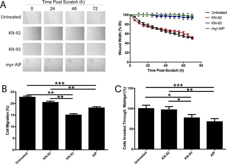
Calcium/calmodulin-stimulated protein kinase II (CaMKII) is a multi-functional kinase that controls a range of cellular functions, including proliferation, differentiation and apoptosis. The biological properties of CaMKII are regulated by multi-site phosphorylation. However, the role that CaMKII phosphorylation plays in cancer cell metastasis has not been examined. We demonstrate herein that CaMKII expression and phosphorylation at T286 is increased in breast cancer when compared to normal breast tissue, and that increased CAMK2 mRNA is associated with poor breast cancer patient prognosis (worse overall and distant metastasis free survival). Additionally, we show that overexpression of WT, T286D and T286V forms of CaMKII in MDA-MB-231 and MCF-7 breast cancer cells increases invasion, migration and anchorage independent growth, and that overexpression of the T286D phosphomimic leads to a further increase in the invasive, migratory and anchorage independent growth capacity of these cells. Pharmacological inhibition of CaMKII decreases MDA-MB-231 migration and invasion. Furthermore, we demonstrate that overexpression of T286D, but not WT or T286V-CaMKII, leads to phosphorylation of FAK, STAT5a, and Akt. These results demonstrate a novel function for phosphorylation of CaMKII at T286 in the control of breast cancer metastasis, offering a promising target for the development of therapeutics to prevent breast cancer metastasis.


Figures

Figure 1
Figure 1. CaMKII phosphorylation at T286 is associated with more aggressive breast cancer, and high CAMK2 expression predicts worse overall and distant metastasis free survival in breast cancer patients.
(A) Total endogenous CaMKII and T286 phosphorylation was determined by western blot. GAPDH expression was used as a loading control. Blots are representative of three independent experiments. (B) The proportion of CaMKII phosphorylated at T286 was determined by normalising the level of T286 phosphorylation to total CaMKII expression. **denotes statistical significance p < 0.01, as determined by one-way ANOVA. (C) Kaplan-Meier curves showing the overall survival [OS] and (D) distant metastasis free survival [DMFS] in a publically available 1881-sample breast cancer data set, with high (blue), mid (red) or low (grey) expression of the CAMK2 family in breast cancer tumours when assessing all tumour subtypes together. P values were computed by a likelihood-ratio test.
Figure 2
Figure 2. CaMKII expression and phosphorylation at T286 is increased in primary breast cancer and lymph node metastases tissues.
(A,D) Normal breast, (B,E) primary breast cancer, and (C,F) lymph node metastases were examined for (A–C) total CaMKII and (D–F) pT286-CaMKII expression by immunohistochemistry. Staining was quantified and expressed as an H-score. (G) Quantification of total CaMKII and (H) pT286-CaMKII expression in 70 primary breast cancer, 40 matched normal breast, and 10 lymph node metastases cores. Photomicrographs are representative of each tissue type. *denotes statistical significance p < 0.05, **p < 0.01, ***p < 0.001, ****p < 0.00001, as determined by one-way ANOVA.
Figure 3
Figure 3. CaMKIIα controls cellular proliferation in breast cancer cells.
(A) MDA-MB-231 cells inducibly expressing or (B) MCF-7 cells stably expressing empty vector (EV), wild-type (WT), T286D, or T286V CaMKII were generated. Cell viability was measured at 0, 24, 48, and 72 h post-CaMKII expression or plating via Cell Titre Blue Assay. *denotes statistical significant difference from EV control cells, as determined by one-way ANOVA (p < 0.05). n = 4. (C) MDA-MB-231 and (D) MCF-7 cells expressing CaMKII mutants were grown for 8 (MDA-MB-231) or 21 (MCF-7) days. After this time, colonies were stained with 0.5% crystal violet/10% methanol/PBS for 30 mins, and then counted. Photomicrographs are representative of three independent experiments, performed in triplicate. *denotes statistical significant difference p < 0.05, as determined by one-way ANOVA.
Figure 4
Figure 4. T286D phosphomimic mutation of CaMKII enhances breast cancer cell migration.
(A) MDA-MB-231 and (B) MCF-7 cells expressing empty vector (EV), wild-type (WT), T286D, or T286V CaMKII were grown to confluence, and a wound was made by scratching with a pipette tip. The wounds were photographed hourly for 72 h to measure wound closure over time. Photomicrographs are representative of three independent experiments performed in triplicate. Wound widths are expressed as % of 0 h wound width. *denotes statistical significance from EV control cells (p < 0.05). ϕdenotes statistical significance compared to WT and T286V cells (p < 0.05), as determined by one-way ANOVA. n = 3. (C) MDA-MB-231 and (D) MCF-7 cells expressing CaMKII mutants were placed in the upper chamber of a Transwell, and allowed to migrate through the uncoated membrane (8 μm pore) for 4 h. n = 3. *denotes statistical significance p < 0.05, ***p < 0.001, ****p < 0.0001 as determined by one-way ANOVA.
Figure 5
Figure 5. T286D phosphomimic mutation of CaMKII enhances breast cancer cell invasion.
(A) MDA-MB-231 and (B) MCF-7 cells expressing CaMKII mutants were examined for ability to invade through Matrigel plugs. n = 3. *denotes statistical significance p < 0.05, **p < 0.01, ***p < 0.001, as determined by one-way ANOVA.
Figure 6
Figure 6. T286D phosphomimic mutation of CaMKII enhances anchorage independent growth.
(A) MDA-MB-231 and (B) MCF-7 cells expressing empty vector (EV), wild-type (WT), T286D, or T286V CaMKII were grown for 14 (MDA-MB-231) or 24 (MCF-7) days in soft agar. After this time, colonies were stained with 0.5% crystal violet/PBS overnight, and then colonies >50 μm were counted using an inverted microscope. Photomicrographs are representative of three independent experiments, performed in triplicate. Results are presented as the number of colonies. *denotes statistical significant difference p < 0.05, **p < 0.01, as determined by one-way ANOVA.
Figure 7
Figure 7. Pharmacological inhibition of CaMKII activity prevents breast cancer cell migration and invasion.
Confluent monolayers of parental MDA-MB-231 cells were treated with 20 μM KN-92 or KN-93, or 10 μM myr-AIP for 30 min, and a wound was made by scratching the monolayer with a pipette tip. (A) Wounds were photographed hourly for 72 h to measure wound closure over time. Photomicrographs are representative of three independent experiments performed in triplicate. Wound widths are expressed as % of 0h wound width. *denotes statistical significance from untreated cells, p < 0.05, as determined by one-way ANOVA. (B) Following this treatment, cells were placed in the upper chamber of a Transwell, and allowed to migrate through the uncoated membrane (8 μm pore) for 4 h. n = 3. **denotes statistical significance p < 0.01, ***p < 0.001, as determined by one-way ANOVA. (C) Parental MDA-MB-231 cells were treated with 40 μM KN-92 or KN-93, or 10 μM myr-AIP for 30 min, and the ability of cells to invade through Matrigel plugs was examined. *denotes statistical significance p < 0.05, **p < 0.01, ***p < 0.001, as determined by one-way ANOVA.
Figure 8
Figure 8. CaMKII overexpression alters the phosphorylation and expression of multiple proteins in breast cancer cells.
(A) Expression and phosphorylation of 44 proteins were examined by Proteome Profile Human Phospho-Kinase Array following inducible overexpression of empty vector (EV), wild-type (WT), T286D phosphomimic mutant, or T286V phosphonull mutant forms of CaMKII in MDA-MB-231 cells. (B) The relative expression and phosphorylation of these proteins were quantitated by densitometric analysis, and expression normalised to that observed in the EV control cells. (C) The expression and phosphorylation of 7 proteins found to be differentially expressed/phosphorylated following T286D phosphomimic mutant overexpression in the array were confirmed by Western blot in MDA-MB-231 and MCF-7 cells. Blots are representative of three independent experiments. The relative expression of these proteins were quantitated by densitometric analysis, and expression normalised to that observed in EV (D) MDA-MB-231 and (E) MCF-7 cells. *denotes statistical significance from EV cells, Φdenotes statistical significance from WT cells, φdenotes significance from T286V expressing cells.

References

    1. Ferlay J. et al. In IARC CancerBase No. 11 (Lyon, 2013).
    1. Kennecke H. et al. Metastatic behaviour of breast cancer subtypes. J Clin Oncol 28, 3271–3277 (2010). - PubMed
    1. Cross B. M., Breitwieser G. E., Reinhardt T. A. & Rao R. Cellular calcium dynamics in lactation and breast cancer: from physiology to pathology. Am J Physiol Cell Physiol 306, C515–C526 (2014). - PMC - PubMed
    1. Stewart T. A., Yapa K. T. & Monteith G. R. Altered calcium signaling in cancer cells. Biochim Biophys Acta 10.1016/j.bbamem.2014.08.016 (Epub ahead of print) (2014). - PubMed
    1. Chen Y. F., Chen Y. T., Chiu W. T. & Shen M. R. Remodeling of calcium signaling in tumor progression. J Biomed Sci 20, 23 (2013). - PMC - PubMed

Publication types

Substances

LinkOut - more resources