Clinical Trial
. 2016 Sep 8:7:12731.
doi: 10.1038/ncomms12731.

Broad activation of latent HIV-1 in vivo

Affiliations
Clinical Trial

Broad activation of latent HIV-1 in vivo

Kirston Barton et al. Nat Commun. .

Abstract

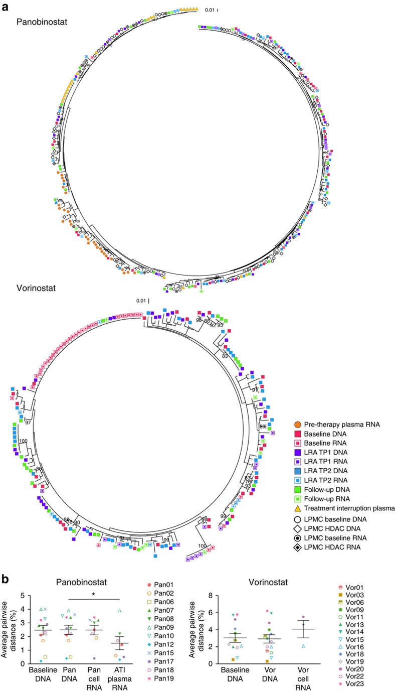
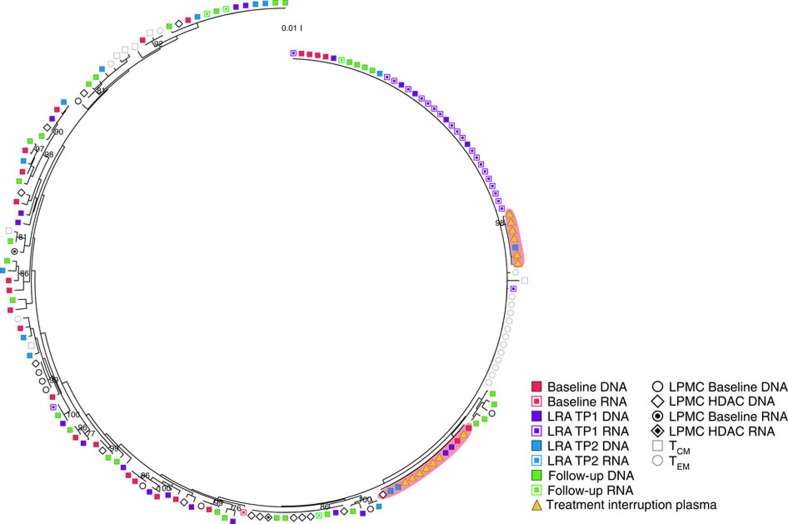
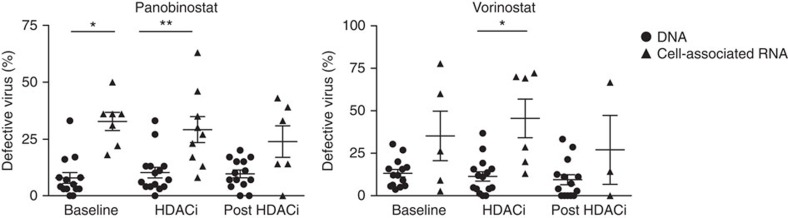
The 'shock and kill' approach to cure human immunodeficiency virus (HIV) includes transcriptional induction of latent HIV-1 proviruses using latency-reversing agents (LRAs) with targeted immunotherapy to purge infected cells. The administration of LRAs (panobinostat or vorinostat) to HIV-1-infected individuals on antiretroviral therapy induces a significant increase in cell-associated unspliced (CA-US) HIV-1 RNA from CD4(+) T cells. However, it is important to discern whether the increases in CA-US HIV-1 RNA are due to limited or broad activation of HIV-1 proviruses. Here we use single-genome sequencing to find that the RNA transcripts observed following LRA administration are genetically diverse, indicating activation of transcription from an extensive range of proviruses. Defective sequences are more frequently found in CA HIV-1 RNA than in HIV-1 DNA, which has implications for developing an accurate measure of HIV-1 reservoir size. Our findings provide insights into the effects of panobinostat and vorinostat as LRAs for latent HIV-1.


Conflict of interest statement

S.R.L. and J.M. received funding from Merck for the investigator-initiated clinical trial of vorinostat described in this manuscript. Payment was made to their institution. The remaining authors declare no competing financial interests.

Figures

Figure 1
Figure 1. Panobinostat and vorinostat non-selectively activate transcription from latent HIV-1 proviruses.
(a) Representative phylogenetic trees of HIV-1 sequences from HIV-infected participants on suppressive ART who received panobinostat (Pan18) or vorinostat (Vor16) showing the genetic relationship of sequences from each time point. For participant Pan18, the plasma samples were collected ∼1 year and 6 months before initiation of antiretroviral therapy and 14 days following the analytical treatment interruption. Peripheral blood samples were collected at baseline, 2 h after the first dose of panobinostat (TP1), 32 days after the first dose of panobinostat (TP2) and 38 days after the final panobinostat dose. Intestinal lamina propria mononuclear cells were collected at baseline (1 week before the first panobinostat dose) and during week 4 of the panobinostat trial. For participant Vor16, peripheral blood samples were collected at baseline, 7 days after the first dose of vorinostat (TP1), 14 days after the first dose of vorinostat (TP2) and 7 days after the final vorinostat dose. (b) Average pairwise distance of cell-associated DNA (Pan n=12, Vor n=12) before and DNA (Pan n=12, Vor n=14) and cell-associated RNA (Pan n= 8, Vor n=3) during vorinostat and panobinostat administration, as well as the plasma HIV-1 RNA following an ATI for the panobinostat trial (n=7). Each data point represents the group mean±s.e.m. The Wilcoxon signed rank test was used to generate the P values. *P≤0.05.
Figure 2
Figure 2. Proviruses in CD4+ T cells contribute to plasma viraemia during an analytical treatment interruption.
Representative phylogenetic tree of HIV-1 sequences (from participant 17) showing that two clusters of identical HIV-1 sequences were detected in the plasma collected during the ATI that are identical to clonal expansions of cell-associated DNA sequences collected during panobinostat administration (red background). The plasma samples were collected 10 and 14 days following the analytical treatment interruption. Peripheral blood samples were collected at baseline, 1 day after the first dose of panobinostat (TP1), 18 days after the first dose of panobinostat (TP2) and 38 days after the final panobinostat dose. Intestinal lamina propria mononuclear cells were collected at baseline (1 week before the first panobinostat dose) and during week 4 of the panobinostat trial. Cell subsets were sorted from peripheral blood samples collected 38 days after the final panobinostat dose.
Figure 3
Figure 3. A significant proportion of cell-associated HIV-1 RNA is defective.
The percentage of defective virus detected in CD4+ T cells collected from HIV-infected participants on ART before, during and following administration of panobinostat (baseline DNA n=14, RNA n=7; on HDACi DNA n=15, RNA n=9; post HDACi DNA n=14, RNA n=6) or vorinostat (baseline DNA n=14, RNA n=5; on HDACi DNA n=15, RNA n=6; post HDACi DNA n=14, RNA n=3). Each data point represents the group mean, and the error bars represent the s.e.m. *P≤0.05, **P≤0.01. The Wilcoxon signed rank test was used to generate the P values.

Similar articles

Cited by

References

    1. Soriano-Sarabia N. et al.. Quantitation of replication-competent HIV-1 in populations of resting CD4+ T cells. J. Virol. 88, 14070–14077 (2014). - PMC - PubMed
    1. Chomont N. et al.. HIV reservoir size and persistence are driven by T cell survival and homeostatic proliferation. Nat. Med. 15, 893–900 (2009). - PMC - PubMed
    1. Wightman F. et al.. Both CD31+ and CD31− naive CD4+ T cells are persistent HIV type 1-infected reservoirs in individuals receiving antiretroviral therapy. J. Infect. Dis. 202, 1738–1748 (2010). - PubMed
    1. Buzon M. J. et al.. HIV-1 persistence in CD4+ T cells with stem cell-like properties. Nat. Med. 20, 139–142 (2014). - PMC - PubMed
    1. Churchill M. J. et al.. Use of laser capture microdissection to detect integrated HIV-1 DNA in macrophages and astrocytes from autopsy brain tissues. J. Neurovirol. 12, 146–152 (2006). - PubMed

Publication types