. 2016 Sep 8:6:33019.
doi: 10.1038/srep33019.

Structural insights into Parkin substrate lysine targeting from minimal Miro substrates

Affiliations

Structural insights into Parkin substrate lysine targeting from minimal Miro substrates

Julian L Klosowiak et al. Sci Rep. .

Abstract

Hereditary Parkinson's disease is commonly caused by mutations in the protein kinase PINK1 or the E3 ubiquitin ligase Parkin, which function together to eliminate damaged mitochondria. PINK1 phosphorylates both Parkin and ubiquitin to stimulate ubiquitination of dozens of proteins on the surface of the outer mitochondrial membrane. However, the mechanisms by which Parkin recognizes specific proteins for modification remain largely unexplored. Here, we show that the C-terminal GTPase (cGTPase) of the Parkin primary substrate human Miro is necessary and sufficient for efficient ubiquitination. We present several new X-ray crystal structures of both human Miro1 and Miro2 that reveal substrate recognition and ubiquitin transfer to be specific to particular protein domains and lysine residues. We also provide evidence that Parkin substrate recognition is functionally separate from substrate modification. Finally, we show that prioritization for modification of a specific lysine sidechain of the cGTPase (K572) within human Miro1 is dependent on both its location and chemical microenvironment. Activation of Parkin by phosphorylation or by binding of pUb is required for prioritization of K572 for modification, suggesting that Parkin activation and acquisition of substrate specificity are coupled.


Figures
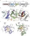
Figure 1
Figure 1. Structure of the hMiro1 EF hand and cGTPase domains.
(a) Bar diagram of hMiro1 domain architecture. The nGTPase (beige) and cGTPase (gray) flank two Ca2+-binding EF hand pairs (blue, EF1; green, EF2); a C-terminal transmembrane domain (M, white) attaches hMiro1 to mitochondria. Parkin-ubiquitinated lysines from all studies to-date are highlighted below. Numbering corresponds to hMiro1 isoform 3; letters (A, B, C) represent hMiro fragments. (b) Crystal structure of hMiro1-BC (aa 177-592). Note three distinct domains: EF1 (blue), EF2 (green), and cGTPase (grey/yellow), joined by linkers Lnk1 (orange), and Lnk2 (red). The structure shown is bound to the non-hydrolyzable GTP analog GMPPCP (CPK representation), with Mg2+ bound at both cEF hands (magenta spheres). (c) Details of Ca2+ coordination by the cEF hands of hMiro1. Ca2+ (yellow sphere) bound at cEF1 and cEF2, revealing pentagonal bipyramidal coordination including critical glutamates E208 and E328. Ca2+-coordinating residues are labeled. hMiro1, human Miro1; cEF, canonical EF hand; hEF, hidden EF hand; LM, ligand mimic.
Figure 2
Figure 2. hMiro1 and hMiro2 are direct substrates of p-S65 Parkin, ubiquitinated on multiple lysines.
(a) hMiro1 is directly ubiquitinated by p-S65 Parkin. 6xHis-tagged full-length hMiro1 (hMiro1-FL, 1 μM) was incubated with various Parkin constructs (0.5 μM): unphosphorylated Parkin (WT), TcPINK1-phosphorylated Parkin (p-WT), and TcPINK1-treated Parkin mutants (S65A, p-C431A), in the presence of E1 (100 nM), E2 (UbcH7, 0.5 μM), and Ub (30 μM). Samples were taken immediately after adding ATP (t = 0 min.) and following incubation at 37 °C (t = 60 min.). Reactions were subjected to immunoblotting to visualize hMiro1-FL (α-His) and Parkin (α-Parkin). (b) hMiro2 is directly ubiquitinated by p-S65 Parkin. Reaction conditions as in (a) but with hMiro2-FL. (c) p-S65 Parkin ubiquitinates hMiro1-FL more efficiently than hMiro2-FL. Time course assay comparing ubiquitination of hMiro1-FL and hMiro2-FL (2 μM) by p-S65 Parkin (0.5 μM). (d) hMiro1-FL and hMiro2-FL are multi-monoubiquitinated by p-S65 Parkin. hMiro1-FL or hMiro2-FL was incubated with p-S65 Parkin in the presence of either wild type (WT) ubiquitin or lysine-less ubiquitin (0 K) incapable of forming ubiquitin chains. hMiro1-FL is efficiently modified at several distinct lysines, as evidenced by the persistence of hMiro1 Ub1–6 bands in the 0 K condition. (e) The positions of all lysine sidechains of hMiro1 are shown in the context of the hMiro1-BC structure (top-view, rotated 90° relative to Fig. 1b). Of 42 lysines in the primary sequence of hMiro1, 12 have been demonstrated to be ubiquitinated (underlined, save K153 of the nGTPase not present in our structure). Two lysines, K235 and K572 (larger font), are found di-Gly modified in our assays.
Figure 3
Figure 3. The cGTPase domain of hMiro is necessary and sufficient for efficient ubiquitination by p-S65 Parkin.
(a) Purified, 6xHis-tagged fragments of hMiro1 (1 μM) were incubated with p-S65 Parkin (0.5 μM) and assessed for ubiquitination (α-His blot). Domains within hMiro1 are labeled as follows: A, corresponding to the nGTPase domain; B, corresponding to the central EF hand region; and C, corresponding to the cGTPase domain (schematized in Fig. 1a). All hMiro1 fragments containing the cGTPase domain were robustly ubiquitinated (hMiro1-FL, hMiro1-BC, hMiro1-C), while the nGTPase alone (hMiro1-A) or the nGTPase with EF hands (hMiro1-BC) showed limited ubiquitination. (b) Fragments of hMiro2 incubated with p-S65 Parkin as in (a). hMiro2 fragments containing the cGTPase domain showed evidence of ubiquitination, albeit at reduced levels as compared to hMiro1. hMiro2-A and hMiro2-AB showed no evidence of ubiquitination. Note that hMiro2 has two common allelic variants, R425 and C425; both variants behaved identically in ubiquitination assays (Supplementary Fig. S12) . Parkin activity was equal throughout (α-Parkin blots).
Figure 4
Figure 4. Structures of hMiro1-C and hMiro2-C reveal cGTPase dimerization via a conserved hydrophobic surface.
(a) Crystal structure of hMiro1-C cGTPase dimer bound to GDP-Pi. Nucleotide binding elements are colored yellow and labeled: P (P-loop); Sw I (Switch I); Sw II (Switch II); G4-G5, guanine nucleotide-binding motifs G4 and G5. Note the two-fold rotational symmetry, highlighted by the mirror image hMiro1 cGTPase label and crystallographic two-fold (arrow). (b) Crystal structure of hMiro2-C cGTPase dimer bound to GDP; labels are as in (a). Note the two-fold rotational symmetry (crystallographic dyad). (c) Location of lysine side chains (black sticks) and hydrophobic residues (orange sticks) at hMiro1-C dimer interface. Lysines with underlined labels were diGly-modified in a dimeric hMiro1-C ubiquitination sample. (d) Location of lysine side chains and hydrophobic residues at hMiro2-C dimer interface. Dimer symmetry is indicated by the mirror image hMiro2 cGTPase label. (e) hMiro1-C and hMiro2-C dimerize in solution via their conserved hydrophobic surfaces. SEC-MALS traces of hMiro2-C (blue, left), hMiro1-C (green, middle), and hMiro1-CM with three mutations in its hydrophobic dimer interface (V418R, Y470S, L472A, orange, right). Both hMiro1-C and hMiro2-C elute at twice their calculated molecular weights (hMiro1-C: 21.9 kD; hMiro2-C: 20.2 kD), while hMiro1-CM elutes as a monomer. Typical SEC differential refractive index (dRI) profiles are shown, normalized for each run (y-axis on the left). In-line MALS profiles across each elution peak are shown (y-axis on the right). Concentration of injected proteins: hMiro1-C (16 mg/mL); hMiro2-C (7 mg/mL); hMiro1-CM (18 mg/mL).
Figure 5
Figure 5. Parkin preferentially targets K572 in the hMiro1 cGTPase domain due to its location and microenvironment.
(a) Disrupting hMiro1-C dimerization alters the cGTPase ubiquitination pattern. Various hMiro1-C (1 μM) constructs were incubated with p-S65 Parkin (0.5 μM) in the presence of either WT Ub or 0 K Ub (30 μM). hMiro1-CD dimer is efficiently ubiquitinated on multiple lysines, as is hMiro1-CD K572R (lanes 1–8). In contrast, the isolated monomeric hMiro1 cGTPase, hMiro1-CM, is primarily monoubiquitinated (lanes 9–12). (b) hMiro1-CM is a mostly single-lysine substrate primarily ubiquitinated at K572. hMiro1-CM Ub1 travels as a doublet characterized by a dark bottom band. A K572R mutation in hMiro1-CM abolishes this dark bottom band, leaving a faint doublet. (c) Structural alignment of the hMiro1 and hMiro2 cGTPases highlights differences in the vicinity of the C-terminal helix. K572 in hMiro1 (blue) corresponds to Q569 in hMiro2 (pink). The chemical environment of the hMiro1 K572 side-chain is also distinct between the two proteins: P553 in hMiro1 vs. A550 in hMiro2, D568 in hMiro1 vs. T565 in hMiro2. (d) A Q569K mutation in hMiro2-C does not increase ubiquitination by p-S65 Parkin as compared to wild-type hMiro2-C. (e) Targeted ubiquitination at hMiro1 K572 is critically sensitive to chemical microenvironment. Mutation of the hMiro1-CM sequence to corresponding residues of hMiro2-C, P553A and D568T, greatly reduce the efficiency and specificity of ubiquitination. The single and double mutants show no preference for K572 targeting, as evidenced by the Ub1 doublet with roughly equal top and bottom band intensities (lanes 6, 8, 10).
Figure 6
Figure 6. Parkin phosphorylation results in both catalytic activation and substrate lysine prioritization.
(a) Phosphorylation of Parkin confers the ability to preferentially target K572 in the hMiro1 cGTPase domain. The indicated Parkin constructs (0.5 μM) were incubated with the minimal hMiro1-CM substrate (1 μM) in the presence of E1, E2, and Ub (30 μM). Untagged S65A Parkin is autoinhibited and shows no activity (lanes 1–4). 6xHis Parkin artificially activated with an N-terminal tag results in low levels of substrate and autoubiquitination (lanes 5–8). The hMiro1-CM Ub1 doublet pattern reflects no apparent preference for ubiquitination at K572, confirmed by the nearly identical pattern in hMiro1-CM K572R (lane 6 vs. 8). In stark contrast, p-S65 Parkin prioritizes K572 ubiquitination over other cGTPase lysines, (dark bottom hMiro1-CM Ub1 doublet band, absent in the hMiro1-CM K572R mutant; lanes 8–12). (b) E2 is dispensable for substrate lysine prioritization by p-S65 Parkin. Reaction scheme as in (a), except E1, E2 and Ub were replaced with Ub chemically activated at its C-terminus (Ub-MES; 100 μM). Ub-MES has a mild activating effect on untagged Parkin, generating a weak hMiro1-CM Ub1 doublet with evidence for K572 ubiquitination, but not prioritization (lanes 1–4). Ub-MES exerts a robust activating effect on artificially disinhibited 6xHis Parkin, but yields an hMiro1-CM ubiquitination pattern qualitatively identical to untagged Parkin (lanes 5–8). Phosphorylated Parkin is able to efficiently target K572 in the absence of E2 (lanes 9–12). (c) Comparison of the effect of pUb on various Parkin constructs. Parkin (0.5 μM) was incubated with hMiro1-CM (1 μM) in the presence of E1, E2, and 30 μM Ub or 20 μM Ub + 10 μM pUb. pUb activates p-S65A Parkin for substrate ubiquitination, autoubiquitination and Ub chain synthesis (lanes 1–4). pUb has a very mild activating effect on artificially disinhibited 6xHis Parkin (lanes 5–8). pUb has a differential effect on p-S65 Parkin, apparently stimulating hMiro1CM ubiquitination while simultaneously suppressing Parkin autoubiquitination and Ub chain synthesis (lanes 9–12).

Similar articles

Cited by

References

    1. Pickrell A. M. & Youle R. J. The Roles of PINK1, Parkin, and Mitochondrial Fidelity in Parkinson’s Disease. Neuron 85, 257–273 (2015). - PMC - PubMed
    1. Kitada T. et al. Mutations in the parkin gene cause autosomal recessive juvenile parkinsonism. Nature 392, 605–608 (1998). - PubMed
    1. Valente E. M. Hereditary Early-Onset Parkinson’s Disease Caused by Mutations in PINK1. Science 304, 1158–1160 (2004). - PubMed
    1. Narendra D., Walker J. E. & Youle R. Mitochondrial Quality Control Mediated by PINK1 and Parkin: Links to Parkinsonism. Cold Spring Harb Perspect Biol 4, a011338–a011338 (2012). - PMC - PubMed
    1. Narendra D. P. et al. PINK1 is selectively stabilized on impaired mitochondria to activate Parkin. PLoS Biol 8, e1000298 (2010). - PMC - PubMed

Publication types

MeSH terms

LinkOut - more resources