

Secreted primary human malignant mesothelioma exosome signature reflects oncogenic cargo
- PMID: 27605433
- PMCID: PMC5015102
- DOI: 10.1038/srep32643
Secreted primary human malignant mesothelioma exosome signature reflects oncogenic cargo
Abstract
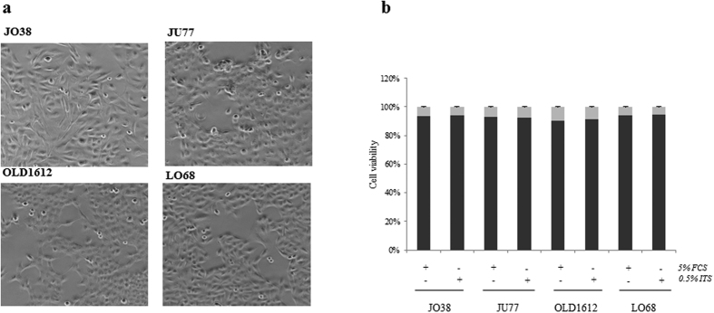
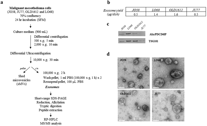
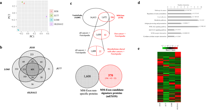
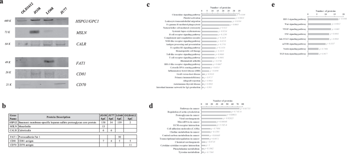
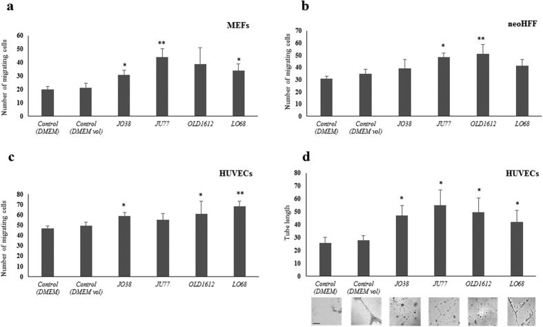
Malignant mesothelioma (MM) is a highly-aggressive heterogeneous malignancy, typically diagnosed at advanced stage. An important area of mesothelioma biology and progression is understanding intercellular communication and the contribution of the secretome. Exosomes are secreted extracellular vesicles shown to shuttle cellular cargo and direct intercellular communication in the tumour microenvironment, facilitate immunoregulation and metastasis. In this study, quantitative proteomics was used to investigate MM-derived exosomes from distinct human models and identify select cargo protein networks associated with angiogenesis, metastasis, and immunoregulation. Utilising bioinformatics pathway/network analyses, and correlation with previous studies on tumour exosomes, we defined a select mesothelioma exosomal signature (mEXOS, 570 proteins) enriched in tumour antigens and various cancer-specific signalling (HPGD/ENO1/OSMR) and secreted modulators (FN1/ITLN1/MAMDC2/PDGFD/GBP1). Notably, such circulating cargo offers unique insights into mesothelioma progression and tumour microenvironment reprogramming. Functionally, we demonstrate that oncogenic exosomes facilitate the migratory capacity of fibroblast/endothelial cells, supporting the systematic model of MM progression associated with vascular remodelling and angiogenesis. We provide biophysical and proteomic characterisation of exosomes, define a unique oncogenic signature (mEXOS), and demonstrate the regulatory capacity of exosomes in cell migration/tube formation assays. These findings contribute to understanding tumour-stromal crosstalk in the context of MM, and potential new diagnostic and therapeutic extracellular targets.
Figures
Similar articles
-
Oncogenic and Non-Malignant Pancreatic Exosome Cargo Reveal Distinct Expression of Oncogenic and Prognostic Factors Involved in Tumor Invasion and Metastasis.Proteomics. 2019 Apr;19(8):e1800158. doi: 10.1002/pmic.201800158. Epub 2019 Apr 9. Proteomics. 2019. PMID: 30893511
-
Proteomic Profiling of Small Extracellular Vesicles Secreted by Human Pancreatic Cancer Cells Implicated in Cellular Transformation.Sci Rep. 2020 May 7;10(1):7713. doi: 10.1038/s41598-020-64718-6. Sci Rep. 2020. PMID: 32382024 Free PMC article.
-
Exosomes: Key mediators of metastasis and pre-metastatic niche formation.Semin Cell Dev Biol. 2017 Jul;67:3-10. doi: 10.1016/j.semcdb.2017.01.004. Epub 2017 Jan 8. Semin Cell Dev Biol. 2017. PMID: 28077297 Review.
-
The Influence of a Stressful Microenvironment on Tumor Exosomes: A Focus on the DNA Cargo.Int J Mol Sci. 2020 Nov 19;21(22):8728. doi: 10.3390/ijms21228728. Int J Mol Sci. 2020. PMID: 33227947 Free PMC article. Review.
-
Exosomal transfer of miR-126 promotes the anti-tumour response in malignant mesothelioma: Role of miR-126 in cancer-stroma communication.Cancer Lett. 2019 Oct 28;463:27-36. doi: 10.1016/j.canlet.2019.08.001. Epub 2019 Aug 7. Cancer Lett. 2019. PMID: 31400405
Cited by
-
Chronic methamphetamine interacts with BDNF Val66Met to remodel psychosis pathways in the mesocorticolimbic proteome.Mol Psychiatry. 2021 Aug;26(8):4431-4447. doi: 10.1038/s41380-019-0617-8. Epub 2019 Dec 10. Mol Psychiatry. 2021. PMID: 31822818
-
Exploring the key communicator role of exosomes in cancer microenvironment through proteomics.Proteome Sci. 2019 Oct 29;17:5. doi: 10.1186/s12953-019-0154-z. eCollection 2019. Proteome Sci. 2019. PMID: 31686989 Free PMC article. Review.
-
Progress in the Management of Malignant Pleural Mesothelioma in 2017.J Thorac Oncol. 2018 May;13(5):606-623. doi: 10.1016/j.jtho.2018.02.021. Epub 2018 Mar 8. J Thorac Oncol. 2018. PMID: 29524617 Free PMC article. Review.
-
Calbindin 2 as a Novel Biomarker and Therapeutic Target for Abdominal Aortic Aneurysm: Integrative Analysis of Human Proteomes and Genetics.J Am Heart Assoc. 2025 May 6;14(9):e039195. doi: 10.1161/JAHA.124.039195. Epub 2025 May 2. J Am Heart Assoc. 2025. PMID: 40314374 Free PMC article.
-
Human Periprostatic Adipose Tissue: Secretome from Patients With Prostate Cancer or Benign Prostate Hyperplasia.Cancer Genomics Proteomics. 2019 Jan-Feb;16(1):29-58. doi: 10.21873/cgp.20110. Cancer Genomics Proteomics. 2019. PMID: 30587498 Free PMC article.
References
Publication types
MeSH terms
Substances
LinkOut - more resources
Full Text Sources
Other Literature Sources
Medical
Research Materials
Miscellaneous
