. 2016 Jul 1;3(8):637-49.
doi: 10.1002/acn3.327. eCollection 2016 Aug.

Tumor prevention facilitates delayed transplant of stem cell-derived motoneurons

Affiliations

Tumor prevention facilitates delayed transplant of stem cell-derived motoneurons

Philippe Magown et al. Ann Clin Transl Neurol. .

Abstract

Objective: Nerve injuries resulting in prolonged periods of denervation result in poor recovery of motor function. We have previously shown that embryonic stem cell-derived motoneurons transplanted at the time of transection into a peripheral nerve can functionally reinnervate muscle. For clinical relevance, we now focused on delaying transplantation to assess reinnervation after prolonged denervation.

Methods: Embryonic stem cell-derived motoneurons were transplanted into the distal segments of transected tibial nerves in adult mice after prolonged denervation of 1-8 weeks. Twitch and tetanic forces were measured ex vivo 3 months posttransplantation. Tissue was harvested from the transplants for culture and immunohistochemical analysis.

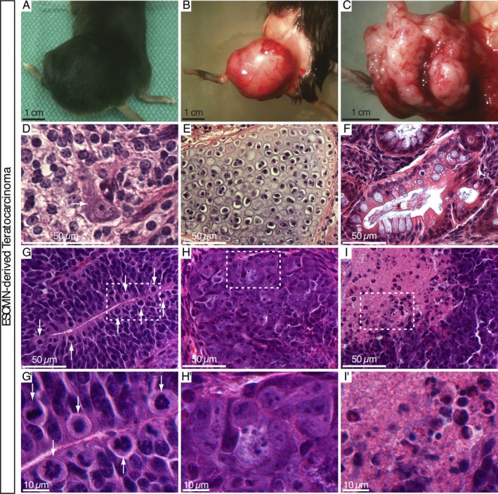
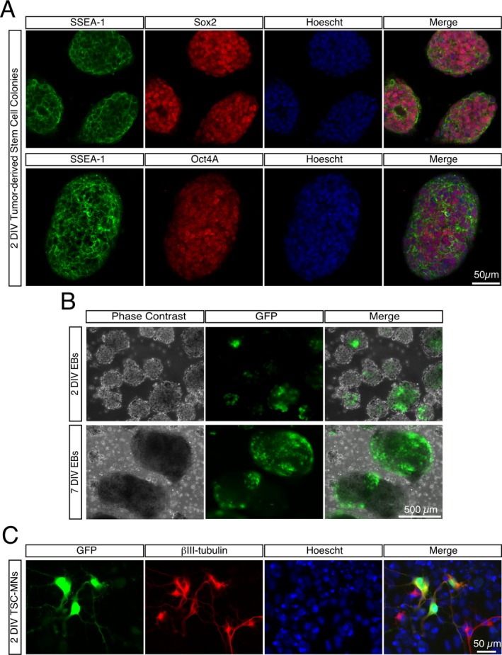
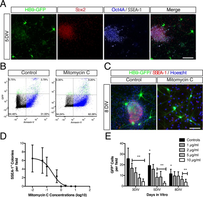
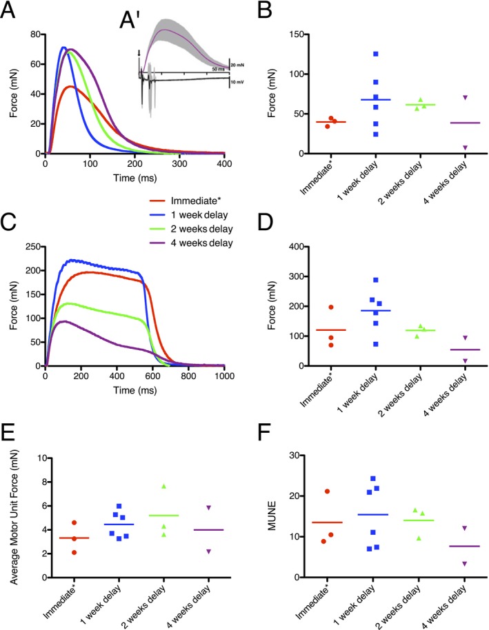
Results: In this delayed reinnervation model, teratocarcinomas developed in about one half of transplants. A residual multipotent cell population (~ 6% of cells) was found despite neural differentiation. Exposure to the alkylating drug mitomycin C eliminated this multipotent population in vitro while preserving motoneurons. Treating neural differentiated stem cells prior to delayed transplantation prevented tumor formation and resulted in twitch and tetanic forces similar to those in animals transplanted acutely after denervation.

Interpretation: Despite a neural differentiation protocol, embryonic stem cell-derived motoneurons still carry a risk of tumorigenicity. Pretreating with an antimitotic agent leads to survival and functional muscle reinnervation if performed within 4 weeks of denervation in the mouse.


Figures

Figure 1
Figure 1
Embryonic stem cell‐derived motoneurons (ESCMN) transplants led to formation of teratocarcinomas. (A–C) Macroscopic appearance of tumor. This first tumor appeared rapidly and unexpectedly. Once aware of this issue, we ensured that no further tumors grew larger than a palpable size of 1 cm. (D–I) Microscopic images of tumors originating from transplanted ESCMNs. All three germ lineages were present in tumors consistent with the formation of a teratomatous tumors: epidermal lineage (neuron: arrow, D), mesodermal lineage (cartilage: E), and endodermal lineage (ciliated glandular epithelium with goblet cells: F). Characteristics of a malignant teratoma (teratocarcinoma): high mitotic rate (arrows pointing at mitotic figures; G, enlarged view G'), hypercellularity with nuclear atypia (H, enlarged view H'), and intratumoral necrosis (I, enlarged view I').
Figure 2
Figure 2
Pluripotent cells isolated from teratocarcinomas generated from transplanted ESCMNs can be differentiated into motoneuron (MNs) in vitro. (A) Pluripotent cells isolated from dissociated teratocarcinoma tissue formed colonies expressing the stem cell markers SSEA‐1, Sox2, and Oct4A. Hoechst staining was used to visualize individual nuclei. (B) Following treatment with retinoic acid and a smoothen agonist, embryonic bodies (EBs) generated from teratocarcinoma‐derived cells contain GFP + MNs after 2 and 7 days in vitro. (C) Dissociated and plated EBs that were treated for 5 days in vitro with retinoic acid and a smoothened agonist, contained GFP + MNs and β III‐tubulin+ cells that were GFP after 2 days in vitro. ESCMNs, embryonic stem cell‐derived motoneurons.
Figure 3
Figure 3
Residual pluripotent cells postembryonic stem cell‐derived motoneurons (ESCMN) differentiation from HBGB6 are sensitive to mitomycin C. (A) Dissociated differentiated embryonic bodies (EBs) grown for 5 days in vitro on matrigel demonstrate the formation of colonies expressing pluripotent markers (SSEA‐1, Oct4A, Sox2) in the absence of LIF and PMEF. Scale bar: 100 μm. (B) FACS sorting of SSEA‐1+ cells from dissociated and annexin‐V immunolabeled EBs (previously treated with retinoic acid and a smoothened agonist) without and with pretreatment with mitomycin C (1 μg/mL for 2 h) 12 h prior to sorting. The shift to the right indicates that the majority of SSEA‐1+ cells expressed annexin‐V (but not GFP) after mitomycin C exposure. (C) Seven days following treatment with 1 μg/mL mitomycin C, dissociated EBs did not contain any colonies of SSEA‐1+ cells (right), but these pluripotent cells were present in untreated EBs (left). Scale bar 100 μm. (D) Dose–response of SSEA‐1+ colonies 7 days in vitro after mitomycin C treatment. No colonies were found when EBs were treated with mitomycin C concentrations of 1 μg/mL or above. *P < 0.05 compared to controls. (E) Toxicity of mitomycin C on ESCMNs showing statistically significant effects with concentrations of 5 μg/mL or greater at all times points compared to controls. **P < 0.01 compared with controls of the same time point, by two‐way ANOVA and Bonferonni multiple comparisons.
Figure 4
Figure 4
Reinnervation after delayed transplantation of embryonic stem cell‐derived motoneurons (ESCMNs) treated with mitomycin C. (A, B) Twitch force and (C, D) 50 Hz tetanic force of the medial gastrocnemius (MG) in delayed transplantation (1, 2, and 4 weeks delay). Traces in A and C are from the same animals. No group showed a statistically significant difference compared to immediate transplants by Kruskal–Wallis test. (A') Four‐week delayed transplant curve force with range (in gray) of response shown in (A) superimposed on EMG signal. Depolarization triplets can be seen in the EMG. Arrow indicates the stimulus artifact. (E) Average MU force from transplanted mice obtained by force gradation with increasing stimulus. (F) Motor unit number estimation. No statistical significant difference between groups. *Forces from immediate transplants are those of HBGB6 ESCMNs not treated with mitomycin C. MU, motor unit.

Similar articles

Cited by

References

    1. Bromberg MB. Quality of life in amyotrophic lateral sclerosis. Phys Med Rehabil Clin N Am 2008;19:591–605. - PubMed
    1. Boakye M, Leigh BC, Skelly AC. Quality of life in persons with spinal cord injury: comparisons with other populations. J Neurosurg Spine 2012;17(1 Suppl):29–37. - PubMed
    1. Carlstedt T, Cullheim S. Spinal cord motoneuron maintenance, injury and repair. Prog Brain Res 2000;127:501–514. - PubMed
    1. Prochazka A, Mushahwar VK, McCreery DB. Neural prostheses. J Physiol (Lond) 2001;533(Pt 1):99–109. - PMC - PubMed
    1. Popovic D, Gordon T, Rafuse VF, Prochazka A. Properties of implanted electrodes for functional electrical stimulation. Ann Biomed Eng 1991;19:303–316. - PubMed

LinkOut - more resources