. 2016 Sep 8:5:e20125.
doi: 10.7554/eLife.20125.

Two locus inheritance of non-syndromic midline craniosynostosis via rare SMAD6 and common BMP2 alleles

Affiliations

Two locus inheritance of non-syndromic midline craniosynostosis via rare SMAD6 and common BMP2 alleles

Andrew T Timberlake et al. Elife. .

Abstract

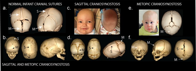
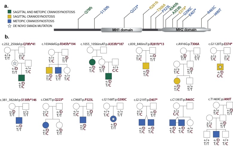
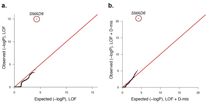
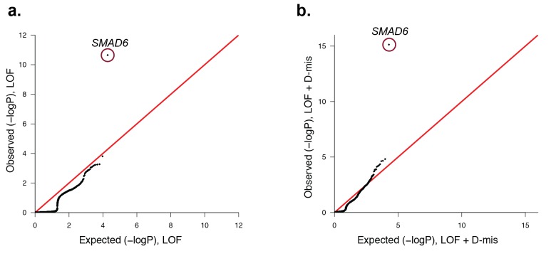
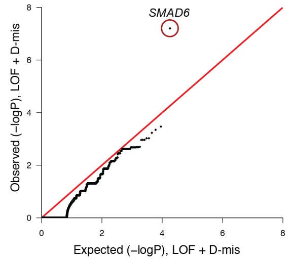
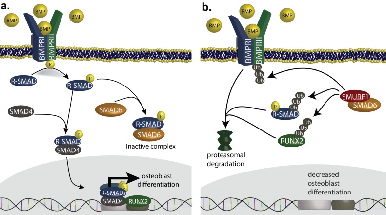
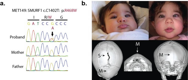
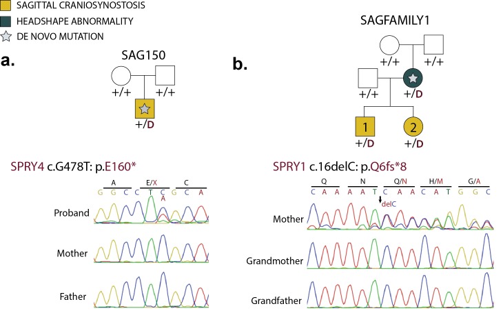
Premature fusion of the cranial sutures (craniosynostosis), affecting 1 in 2000 newborns, is treated surgically in infancy to prevent adverse neurologic outcomes. To identify mutations contributing to common non-syndromic midline (sagittal and metopic) craniosynostosis, we performed exome sequencing of 132 parent-offspring trios and 59 additional probands. Thirteen probands (7%) had damaging de novo or rare transmitted mutations in SMAD6, an inhibitor of BMP - induced osteoblast differentiation (p<10-20). SMAD6 mutations nonetheless showed striking incomplete penetrance (<60%). Genotypes of a common variant near BMP2 that is strongly associated with midline craniosynostosis explained nearly all the phenotypic variation in these kindreds, with highly significant evidence of genetic interaction between these loci via both association and analysis of linkage. This epistatic interaction of rare and common variants defines the most frequent cause of midline craniosynostosis and has implications for the genetic basis of other diseases.

Keywords: BMP2; SMAD6; SMURF1; SPRY1; SPRY4; chromosomes; craniofacial; craniosynostosis; de novo mutation; evolutionary biology; exome sequencing; genes; genomics; human; human genetics; incomplete penetrance.


Conflict of interest statement

The authors declare that no competing interests exist.

Figures

Figure 1.
Figure 1.. Phenotypes of midline craniosynostosis.
(a) Normal infant skull with patent sagittal (S) and metopic (M) sutures. (b) Three-dimensional reconstruction of computed tomography (3D CT) demonstrating premature fusion of both the sagittal and metopic sutures. (c) A three-month-old boy with sagittal craniosynostosis featuring scaphocephaly (narrow and elongated cranial vault), and frontal bossing. (d) 3D CT reconstruction of a one-month-old boy found to have sagittal craniosynostosis. (e) A six-month-old boy presenting with trigonocephaly (triangulation of the cranial vault, with prominent forehead ridge resulting from premature fusion of the metopic suture) and hypotelorism (abnormally decreased intercanthal distance, also a result of premature fusion of the metopic suture). 3D CT reconstruction demonstrated metopic craniosynostosis. (f) 3D CT reconstruction demonstrating premature fusion of the metopic suture with characteristic trigonocephaly and hypotelorism. DOI: http://dx.doi.org/10.7554/eLife.20125.003
Figure 2.
Figure 2.. Segregation of SMAD6 mutations and BMP2 SNP genotypes in pedigrees with midline craniosynostosis.
(a) Domain structure of SMAD6 showing location of the MH1 and MH2 domains. The MH1 domain mediates DNA binding and negatively regulates the functions of the MH2 domain, while the MH2 domain is responsible for transactivation and mediates phosphorylation-triggered heteromeric assembly with receptor SMADs. De novo or rare damaging mutations identified in craniosynostosis probands are indicated. Color of text denotes suture(s) showing premature closure. (b) Pedigrees harboring de novo (denoted by stars within pedigree symbols) or rare transmitted variants in SMAD6. Filled and unfilled symbols denote individuals with and without craniosynostosis, respectively. The SMAD6 mutation identified in each kindred is noted above each pedigree. Below each symbol, genotypes are shown first for SMAD6 (with 'D' denoting the damaging allele) and for rs1884302 risk locus downstream of BMP2, (with 'T' conferring protection from and 'C' conferring increased risk of craniosynostosis). All 17 subjects with craniosynostosis have SMAD6 mutations, and 14/17 have also inherited the risk allele at rs1884302, whereas only 3 of 16 SMAD6 mutation carriers without the rs1884302 risk allele have craniosynostosis. DOI: http://dx.doi.org/10.7554/eLife.20125.006
Figure 2—figure supplement 1.
Figure 2—figure supplement 1.. Plots of independent Illumina sequencing reads in a parent-offspring trio showing de novo SMAD6 mutation.
The reference sequence of a segment of SMAD6 that includes base 15:67073502 (denoted by arrow) is shown in the top row, with red, blue, green and yellow squares representing A, C, G, T, respectively. Below, all independent reads that map to this interval are shown. The results show that the proband has 23 reads of reference ‘G’, and 10 reads of non-reference ‘T’. Only the reference ‘G’ is seen in both parents, providing evidence of a de novo mutation. DOI: http://dx.doi.org/10.7554/eLife.20125.009
Figure 2—figure supplement 2.
Figure 2—figure supplement 2.. Confirmation of SMAD6 mutations by Sanger sequencing of PCR products.
Sanger sequencing traces of PCR amplicons containing SMAD6 mutations identified by exome sequencing are shown. Above each trace or set of traces, the kindred ID, mutation identified in the DNA sequence and its impact on SMAD6 protein is indicated. Above sequence traces, the inferred DNA sequence is shown, along with the inferred amino acid sequence (shown in single letter code). Heterozygous mutations are indicated beneath the wild-type sequence and non-reference amino acid sequences are shown in red. Deleted and inserted bases are denoted, and result in an overlap of wild-type and mutant sequences. DOI: http://dx.doi.org/10.7554/eLife.20125.010
Figure 3.
Figure 3.. Quantile-quantile plots of observed versus expected p-values comparing the burden of rare LOF and damaging (LOF + D-mis) variants in protein-coding genes in craniosynostosis cases.
Rare (allele frequency <2 × 10⁻⁵ in the ExAC03 database) loss of function (LOF) and damaging missense (D-mis) variants were identified in 191 probands. The probability of the observed number of variants in each gene occurring by chance was calculated from the total number of observed variants and the length of the coding region of each gene using the binomial test. The distribution of observed P-values compared to the expected distribution is shown. (a) Q-Q plot for rare LOF variants in each gene from a total of 1135 LOF variants identified in probands. The distribution of observed p-values closely conforms to expectation with the exception of SMAD6, which shows p=1.1 × 10⁻¹⁵ and 156-fold enrichment in cases. (b) Q-Q plot for rare damaging (LOF + D-mis) variants in each gene from a total of 3156 damaging variants in probands. Again, SMAD6 deviates greatly from the expected distribution, with p<10–20 and 91-fold enrichment. DOI: http://dx.doi.org/10.7554/eLife.20125.012
Figure 3—figure supplement 1.
Figure 3—figure supplement 1.. Quantile-quantile plots comparing all transmitted, damaging variants in protein-coding genes in 191 probands with midline craniosynostosis to the expected binomial distribution.
De novo variants were excluded from this analysis, leaving 1122 rare (ExAC allele frequency < 2 x10⁻⁵), transmitted LOF variants and 3115 transmitted damaging (LOF + D-mis) variants. All genes closely matched expectation, with the exception of SMAD6. (a) There were 6 transmitted SMAD6 LOF mutations, a 118-fold enrichment compared to the expected 0.05 (p=2.2 × 10⁻¹¹). (b) Similarly, there were 10 transmitted damaging SMAD6 variants, a 71-fold enrichment compared to the expected 0.14 (p=7.0 × 10⁻¹⁶). The results demonstrate genome-wide significance of rare transmitted variants in SMAD6 independent of de novo mutations. DOI: http://dx.doi.org/10.7554/eLife.20125.014
Figure 3—figure supplement 2.
Figure 3—figure supplement 2.. Principal-component analysis of 191 probands and 3337 European autism controls.
(a) Principal component analysis of exome sequence genotypes from 191 probands with sagittal, metopic, or combined sagittal and metopic craniosynostosis clustered along with HapMap subjects. Results identify 172 craniosynostosis subjects that cluster with HapMap European subjects. (b) Principal component analysis of genotypes from exome sequencing data of European autism parent controls (n = 3337) showing clustering with HapMap subjects. In both panels, subjects considered to be of European ancestry are circled. DOI: http://dx.doi.org/10.7554/eLife.20125.015
Figure 3—figure supplement 3.
Figure 3—figure supplement 3.. Quantile-quantile plot of observed versus expected p-values comparing the burden of damaging (LOF + D-mis) variants in protein-coding genes in craniosynostosis cases and controls.
The frequency of rare (allele frequency < 2 × 10⁻⁵ in the ExAC03 database) loss of function and D-mis variants in each gene was compared in 172 European probands with midline craniosynostosis and 3337 European controls. The distribution of observed p-values conforms to expectation with the exception of SMAD6, which deviates significantly from expectation. Because exon 1 of SMAD6 was poorly captured using the V2 capture reagent (used in control samples), 3 damaging variants in exon 1 in cases were excluded from this analysis. DOI: http://dx.doi.org/10.7554/eLife.20125.016
Figure 4.
Figure 4.. SMAD6 inhibits osteoblast differentiation by inhibiting BMP-mediated SMAD signaling (Salazar et al., 2016).
(a) BMP ligands activate BMP receptors, leading to phosphorylation of receptor-regulated SMADs (R-SMADs), which complex with SMAD4 and enter the nucleus, cooperating with RUNX2 to induce osteoblast differentiation. SMAD6 inhibits this signal by competing with SMAD4 for binding to R-SMADs, preventing nuclear translocation. (b) SMAD6 also cooperates with SMURF1, an E3 ubiquitin ligase, to induce ubiquitin-mediated proteasomal degradation of R-SMADs, BMP receptor complexes, and RUNX2. DOI: http://dx.doi.org/10.7554/eLife.20125.019
Figure 5.
Figure 5.. A de novo variant identified in SMURF1.
(a) Sanger sequence electropherogram of a PCR product amplified from the genomic DNA of a proband with metopic craniosynostosis, confirming a de novo R468W mutation in SMURF1, a SMAD6 binding partner. (b) Patient photographs of the proband, who presented with trigonocephaly and mild orbital abnormalities. 3D CT reconstruction demonstrates metopic craniosynostosis, trigonocephaly, and a patent sagittal suture. DOI: http://dx.doi.org/10.7554/eLife.20125.020
Figure 6.
Figure 6.. De novo loss-of-function mutations in Sprouty genes.
(a) Pedigree and Sanger sequencing traces for kindred SAG150, demonstrating a de novo nonsense mutation in SPRY4 (p.E160*) in the proband. (b) Pedigree and Sanger sequencing traces in a kindred with a de novo SPRY1 frameshift mutation (p.Q6fs*8) that was transmitted to two affected offspring. DOI: http://dx.doi.org/10.7554/eLife.20125.022

Comment in

  • doi: 10.7554/eLife.21162

References

    1. Bier E, De Robertis EM. Embryo development. BMP gradients: A paradigm for morphogen-mediated developmental patterning. Science. 2015;348:e20125. doi: 10.1126/science.aaa5838. - DOI - PubMed
    1. Boyadjiev SA, International Craniosynostosis Consortium Genetic analysis of non-syndromic craniosynostosis. Orthodontics & Craniofacial Research. 2007;10:129–137. doi: 10.1111/j.1601-6343.2007.00393.x. - DOI - PubMed
    1. Connerney J, Andreeva V, Leshem Y, Mercado MA, Dowell K, Yang X, Lindner V, Friesel RE, Spicer DB. Twist1 homodimers enhance FGF responsiveness of the cranial sutures and promote suture closure. Developmental Biology. 2008;318:323–334. doi: 10.1016/j.ydbio.2008.03.037. - DOI - PMC - PubMed
    1. Dathe K, Kjaer KW, Brehm A, Meinecke P, Nürnberg P, Neto JC, Brunoni D, Tommerup N, Ott CE, Klopocki E, Seemann P, Mundlos S. Duplications involving a conserved regulatory element downstream of BMP2 are associated with brachydactyly type A2. American Journal of Human Genetics. 2009;84:483–492. doi: 10.1016/j.ajhg.2009.03.001. - DOI - PMC - PubMed
    1. De Rubeis S, He X, Goldberg AP, Poultney CS, Samocha K, Cicek AE, Kou Y, Liu L, Fromer M, Walker S, Singh T, Klei L, Kosmicki J, Shih-Chen F, Aleksic B, Biscaldi M, Bolton PF, Brownfeld JM, Cai J, Campbell NG, Carracedo A, Chahrour MH, Chiocchetti AG, Coon H, Crawford EL, Curran SR, Dawson G, Duketis E, Fernandez BA, Gallagher L, Geller E, Guter SJ, Hill RS, Ionita-Laza J, Jimenz Gonzalez P, Kilpinen H, Klauck SM, Kolevzon A, Lee I, Lei I, Lei J, Lehtimäki T, Lin CF, Ma'ayan A, Marshall CR, McInnes AL, Neale B, Owen MJ, Ozaki N, Parellada M, Parr JR, Purcell S, Puura K, Rajagopalan D, Rehnström K, Reichenberg A, Sabo A, Sachse M, Sanders SJ, Schafer C, Schulte-Rüther M, Skuse D, Stevens C, Szatmari P, Tammimies K, Valladares O, Voran A, Li-San W, Weiss LA, Willsey AJ, Yu TW, Yuen RK, Cook EH, Freitag CM, Gill M, Hultman CM, Lehner T, Palotie A, Schellenberg GD, Sklar P, State MW, Sutcliffe JS, Walsh CA, Scherer SW, Zwick ME, Barett JC, Cutler DJ, Roeder K, Devlin B, Daly MJ, Buxbaum JD, DDD Study. Homozygosity Mapping Collaborative for Autism. UK10K Consortium Synaptic, transcriptional and chromatin genes disrupted in autism. Nature. 2014;515:209–215. doi: 10.1038/nature13772. - DOI - PMC - PubMed