. 2016 Sep 8;11(9):e0161229.
doi: 10.1371/journal.pone.0161229. eCollection 2016.

A Newly Defined and Xeno-Free Culture Medium Supports Every-Other-Day Medium Replacement in the Generation and Long-Term Cultivation of Human Pluripotent Stem Cells

Affiliations

A Newly Defined and Xeno-Free Culture Medium Supports Every-Other-Day Medium Replacement in the Generation and Long-Term Cultivation of Human Pluripotent Stem Cells

Behnam Ahmadian Baghbaderani et al. PLoS One. .

Abstract

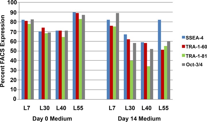
Human pluripotent stem cells (hPSCs) present an unprecedented opportunity to advance human health by offering an alternative and renewable cell resource for cellular therapeutics and regenerative medicine. The present demand for high quality hPSCs for use in both research and clinical studies underscores the need to develop technologies that will simplify the cultivation process and control variability. Here we describe the development of a robust, defined and xeno-free hPSC medium that supports reliable propagation of hPSCs and generation of human induced pluripotent stem cells (hiPSCs) from multiple somatic cell types; long-term serial subculturing of hPSCs with every-other-day (EOD) medium replacement; and banking fully characterized hPSCs. The hPSCs cultured in this medium for over 40 passages are genetically stable, retain high expression levels of the pluripotency markers TRA-1-60, TRA-1-81, Oct-3/4 and SSEA-4, and readily differentiate into ectoderm, mesoderm and endoderm. Importantly, the medium plays an integral role in establishing a cGMP-compliant process for the manufacturing of hiPSCs that can be used for generation of clinically relevant cell types for cell replacement therapy applications.


Conflict of interest statement

All authors are current or previous employees of Lonza, a pharmaceutical company that develops and sells a wide range of products, including cell-based products for research and pharmaceutical use. Lonza may derive benefit from the sale of a product derived from this research. However, this does not alter our adherence to all the PLOS ONE policies on sharing data and materials.

Figures

Fig 1
Fig 1. Differences observed in the stability of distinct hPSC medium formulations.
Four hPSC medium formulations were found to support the maintenance of hPSC lines in culture. To evaluate the stability of each formulation, both freshly supplemented medium and medium that was supplemented and then stored at 4°C for 14 days prior to addition to the hPSC lines, were compared. In this representative example using WA09 cells, all formulations sustained high expression levels of pluripotency markers when the medium was used within seven days after supplementation with bFGF and other factors. However, only the medium containing the L7 supplement formulation was able to sustain robust expression of the hPSC markers with aged medium (14–21 days) post supplementation.
Fig 2
Fig 2. Medium containing thermostable bFGF supports every-other-day supplementation of hPSC cultures.
hPSCs (WA09 hESC line) were seeded at 2 x 10⁵ cells per well in a 6-well tissue culture treated coated with L7 hPSC matrix in the defined hPSC medium. Three days post-seeding, the medium of metabolically active hPSC cultures was replaced with freshly supplemented basal medium containing either bFGF or TS bFGF. Twenty four and 48 hours post-addition, a sample of the medium was taken and the concentration of bFGF present determined by ELISA. A) bFGF concentrations present in the hPSC medium at the time of addition, 24 hours- and 48 hours-post addition (n = 3). B) TS bFGF concentrations present in the hPSC medium at the time of addition, 24 hours- and 48 hours-post addition (n = 3).
Fig 3
Fig 3. Comparison of hPSC morphology and growth using the defined, xeno-free hPSC medium.
Three hPSC lines were adapted to the defined, xeno-free medium formulation supplemented with either bFGF and heparin or TS bFGF without heparin. Cultures seeded at 2 x 10⁴ cells/cm2, received either daily medium changes with medium containing heparin and bFGF or received medium containing TS bFGF but no heparin every other day (EOD). Scale bar: 200 μm.
Fig 4
Fig 4. hPSCs cultured in the defined, xeno-free EOD hPSC medium retain their stem cell characteristics.
Immunodetection of pluripotency antigens in hPSC lines cultivated for 40 passages in the defined, xeno-free L7 hPSC medium with EOD medium changes. Detection of Oct-3/4 and Nanog are shown (red) along with SSEA-4 and TRA-1-60 (green). Individual cell nuclei were visualized using DAPI (blue). Scale bar: 200 μm.
Fig 5
Fig 5. Expression of pluripotency markers in hPSCs cultured for 40 passages using the defined, xeno-free EOD hPSC medium.
Three hPSC lines (two replicates per line) were evaluated for their ability to sustain the expression of markers of pluripotency at passage 10, passage 25 and passage 40. Each hPSC line was independently analyzed by flow cytometry for the expression of SSEA-4, TRA-1-60 and TRA-1-81. Results within each passage were combined and the mean value of marker expression determined. Bars represent mean percentage values. Error bars represent the standard deviation of the mean for each passage.
Fig 6
Fig 6. hPSCs cultured for over 40 passages using the defined, xeno-free hPSC medium differentiate readily to early ectoderm, mesoderm and endoderm (EB formation).
A) Phase contrast images of the hPSC lines just prior to differentiation. B) Embryoid body formation in cultures differentiated for 14 days in culture. C) Antibodies detecting Beta-III-Tubulin (TUJ1) are shown in red; Smooth Muscle Actin (SMA) and Alpha-Feto Protein (AFP) antigens are shown in green. Nuclei were visualized using DAPI (blue). Scale bar: 200 μm.
Fig 7
Fig 7. Comparison of the defined, xeno-free EOD L7 hPSC EOD medium to mTeSR1 E8 medium.
Human hESC line WA07 was serially subcultured for five passages. The number of viable cells per passage were compared by initially seeding 2×10⁴ viable cells per cm2 into three wells of a six-well plate for each medium, using respective passaging solution and matrix. Panel A shows cell attachment and growth of WA07 hESCs at passage 1 in L7 hPSC, mTeSR1TM, and E8TM media. Panel B shows cell attachment and growth of WA07 hESCs at the end of passage 5 in L7 hPSC, mTeSR1, and E8 media. Panel C Panel A shows the viability and total viable cells of WA07 hESCs grown in L7 hPSC, mTeSR1, and E8 media, demonstrating comparative growth for the cells grown in each media. Panel D shows immunocytochemistry analysis of WA07 hESCs grown in L7 hPSC, mTeSR1, and E8 media, demonstrating comparative expression of OCT4 (red), Nanog (red), SSEA4 (green), TRA1-60 (green), and TRA1-81 (green) in each media. Following differentiation of hPSCs into embryoid bodies (Panel E), differentiated WA07 hESCs readily expressed the markers for early ectoderm (detected TUJ1, green), endoderm (detected Alpha-Feto Protein (AFP), green), and mesoderm (detected Smooth Muscle Actin (SMA), green) in L7 hPSC and E8TM media. Cell nuclei are shown by DAPI (blue).

Similar articles

Cited by

References

    1. Guenther MG, Frampton GM, Soldner F, Hockemeyer D, Mitalipova M, Jaenisch R, et al. (2010) Chromatin structure and gene expression programs of human embryonic and induced pluripotent stem cells. Cell Stem Cell 7: 249–257. 10.1016/j.stem.2010.06.015 - DOI - PMC - PubMed
    1. Newman AM, Cooper JB (2010) Lab-specific gene expression signatures in pluripotent stem cells. Cell Stem Cell 7: 258–262. 10.1016/j.stem.2010.06.016 - DOI - PubMed
    1. Ward CM, Barrow KM, Stern PL (2004) Significant variations in differentiation properties between independent mouse ES cell lines cultured under defined conditions. Exp Cell Res 293: 229–238. - PubMed
    1. Guha P, Morgan JW, Mostoslavsky G, Rodrigues NP, Boyd AS (2013) Lack of immune response to differentiated cells derived from syngeneic induced pluripotent stem cells. Cell Stem Cell 12: 407–412. 10.1016/j.stem.2013.01.006 - DOI - PubMed
    1. Amit M, Shariki C, Margulets V, Itskovitz-Eldor J (2004) Feeder layer- and serum-free culture of human embryonic stem cells. Biol Reprod 70: 837–845. - PubMed