. 2016 Sep 8;11(9):e0162518.
doi: 10.1371/journal.pone.0162518. eCollection 2016.

Single-Step Fabrication of Computationally Designed Microneedles by Continuous Liquid Interface Production

Affiliations

Single-Step Fabrication of Computationally Designed Microneedles by Continuous Liquid Interface Production

Ashley R Johnson et al. PLoS One. .

Abstract

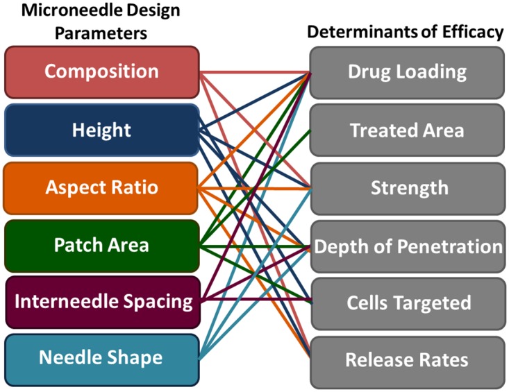
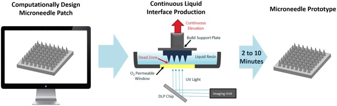
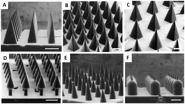
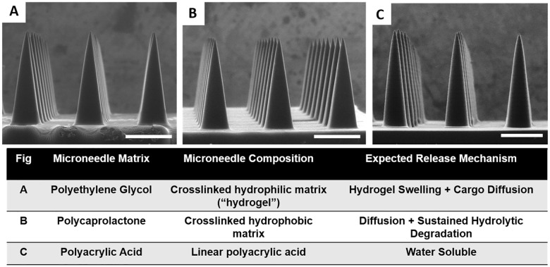
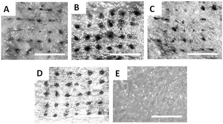
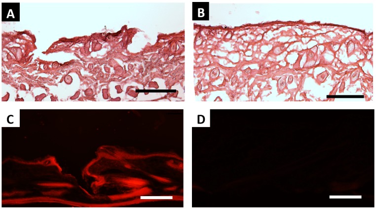
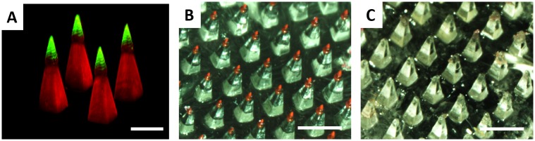
Microneedles, arrays of micron-sized needles that painlessly puncture the skin, enable transdermal delivery of medications that are difficult to deliver using more traditional routes. Many important design parameters, such as microneedle size, shape, spacing, and composition, are known to influence efficacy, but are notoriously difficult to alter due to the complex nature of microfabrication techniques. Herein, we utilize a novel additive manufacturing ("3D printing") technique called Continuous Liquid Interface Production (CLIP) to rapidly prototype sharp microneedles with tuneable geometries (size, shape, aspect ratio, spacing). This technology allows for mold-independent, one-step manufacturing of microneedle arrays of virtually any design in less than 10 minutes per patch. Square pyramidal CLIP microneedles composed of trimethylolpropane triacrylate, polyacrylic acid and photopolymerizable derivatives of polyethylene glycol and polycaprolactone were fabricated to demonstrate the range of materials that can be utilized within this platform for encapsulating and controlling the release of therapeutics. These CLIP microneedles effectively pierced murine skin ex vivo and released the fluorescent drug surrogate rhodamine.


Conflict of interest statement

I have read the journal's policy and the authors of this manuscript have the following competing interests: JRT, DS, AE, and JMD all have an equity stake in Carbon, Inc., which is a venture-backed startup company. This does not alter our adherence to PLOS One policies on data sharing and materials.

Figures

Fig 1
Fig 1. Relationship between microneedle design parameters and therapeutic efficacy.
The left column is a list of microneedle design parameters, whereas the right column is a list of factors affecting the efficacy of microneedles used for transdermal drug delivery. Connections between input design parameters and their effect on efficacy are marked with solid lines.
Fig 2
Fig 2. Continuous Liquid Interface Production (CLIP) Process.
A microneedle patch is computationally designed using a CAD file. The microneedle is then fabricated using CLIP to produce a microneedle prototype within two to ten minutes. CLIP generates the microneedle patch through photopolymerization of a liquid, photoreactive resin using light reflecting off of a DLP chip. Continuous (rather than layer-by-layer) fabrication of the patch is enabled by a “dead-zone” created through oxygen mediated inhibition of the photopolymerization reaction at the window surface. A microneedle patch of virtually any design is created in two to ten minutes.
Fig 3
Fig 3. TMPTA Microneedles of Different Heights.
TMPTA microneedles measuring approximately A) 1000μm C) 700μm and D) 400μm in height with AR = 3. B) Representative image of a microneedle tip with a tip radius of approximately 2.3μm. Scale bars measure 500μm (A, C-D) and 5μm (B), respectively. All patches were generated in less than 90 seconds.
Fig 4
Fig 4. TMPTA Microneedles of Different Shapes.
A) TMPTA microneedles of aspect ratio 2, 3, and 4 (left to right). 1000μm tall TMPTA microneedles with spacing of B) 0.5 base widths and C) 1.5 base widths. Complex microneedle geometries such as D) Arrowhead microneedles E) Tiered microneedles and F) Turret microneedles may improve mechanics of insertion into the skin. Scale bars measure 500μm.
Fig 5
Fig 5. Biocompatible Microneedles.
ESEM images of A) Polyethylene glycol B) Polycaprolactone and C) Polyacrylic acid microneedles measuring approximately 1000μm in height and 333μm in width. Starting resin, resulting microneedle composition, and expected cargo release mechanism for biocompatible microneedles are also provided. Scale bars measure 500μm.
Fig 6
Fig 6. Skin Insertion Tests.
Sites of skin penetration from CLIP Microneedle arrays made of A) PCL B) TMPTA C) PEG and D) Polyacrylic acid on murine skin can be visualized using a tissue marking dye. E) No insertion sites are visualized on a piece of control skin to which no microneedles were applied. Scale bars measure 1mm.
Fig 7
Fig 7. Ex-Vivo Skin Penetration and Dye Release.
H&E stained skin sections show A) epidermal breach upon application of PAA microneedles but B) no epidermal breach in untreated control. C) The application of rhodamine containing polyacrylic acid microneedles releases rhodamine into the skin. D) No fluorescence is visualized in sections to which no microneedles were applied. All scale bars measure 100μm.
Fig 8
Fig 8. Tip loaded microneedles enable complete cargo delivery.
A) Confocal micrograph of a Janus microneedle, where the microneedle base is composed of polycaprolactone encapsulating rhodamine and the microneedle tip is composed of polyacrylic acid encapsulating fluorescein. The rhodamine channel is given in red and the fluorescein channel is given in green; the overlay is displayed in yellow. Janus microneedle with a water soluble rhodamine containing tip B) before and C) after application to murine skin. Scale bars measure 500μm (A) and 1mm (B-C)

References

    1. Prausnitz MR. Microneedles for transdermal drug delivery. Adv Drug Deliv Rev 2004; 56:581–587. - PubMed
    1. Kim YC, Park JH, Prausnitz MR. Microneedles for drug and vaccine delivery. Adv Drug Deliv Rev 2012; 64:1547–1568. 10.1016/j.addr.2012.04.005 - DOI - PMC - PubMed
    1. Haq MI, Smith E, John DN, Kalavala M, Edwards C, Anstey A et al. Clinical administration of microneedles: skin puncture, pain and sensation. Biomed Microdevices 2009; 11:35–47. 10.1007/s10544-008-9208-1 - DOI - PubMed
    1. Ling MH, Chen MC. Dissolving polymer microneedle patches for rapid and efficient transdermal delivery of insulin to diabetic rats. Acta Biomater 2013; 9:8952–8961. 10.1016/j.actbio.2013.06.029 - DOI - PubMed
    1. DeMuth PC, Min Y, Huang B, Kramer JA, Miller AD, Barouch DH et al. Polymer multilayer tattooing for enhanced DNA vaccination. Nat Mater 2013; 12:367–376. 10.1038/nmat3550 - DOI - PMC - PubMed