. 2016 Sep 8;11(9):e0162186.
doi: 10.1371/journal.pone.0162186. eCollection 2016.

Alpha1-Antitrypsin Attenuates Renal Fibrosis by Inhibiting TGF-β1-Induced Epithelial Mesenchymal Transition

Affiliations

Alpha1-Antitrypsin Attenuates Renal Fibrosis by Inhibiting TGF-β1-Induced Epithelial Mesenchymal Transition

Jang-Hee Cho et al. PLoS One. .

Abstract

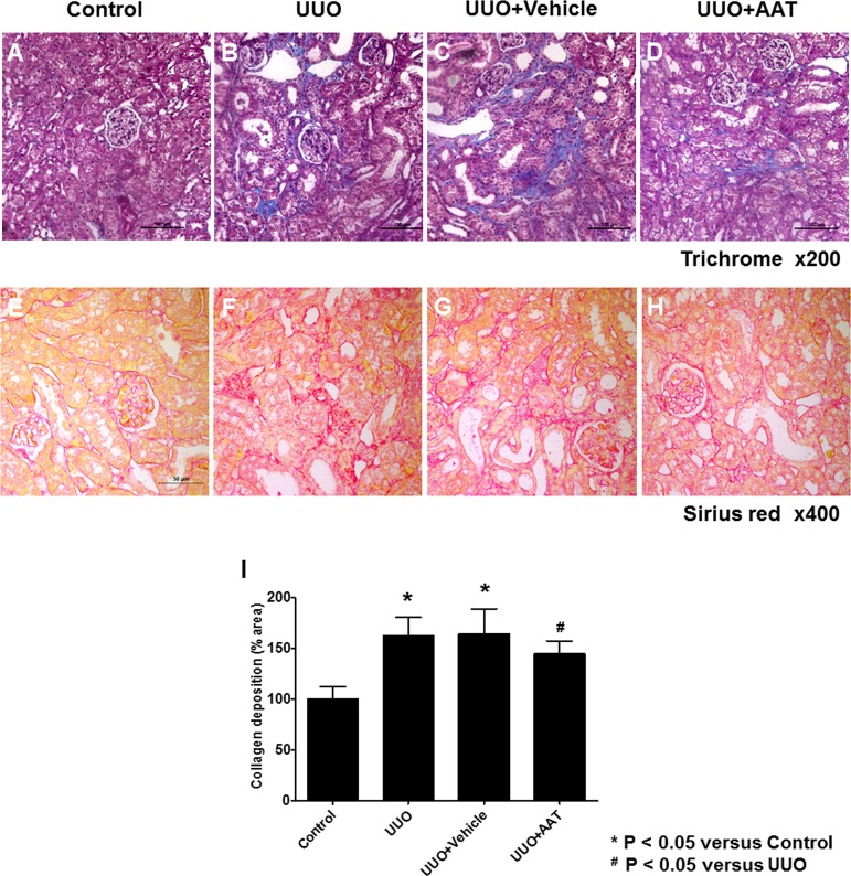
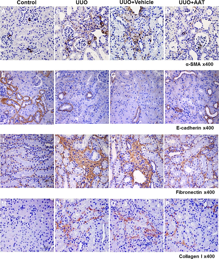
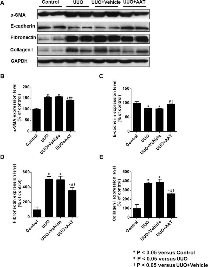
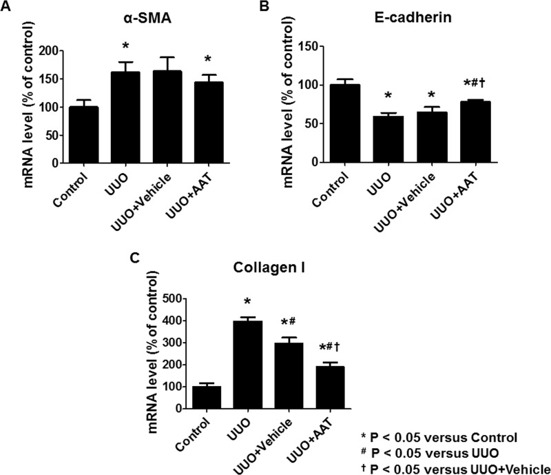
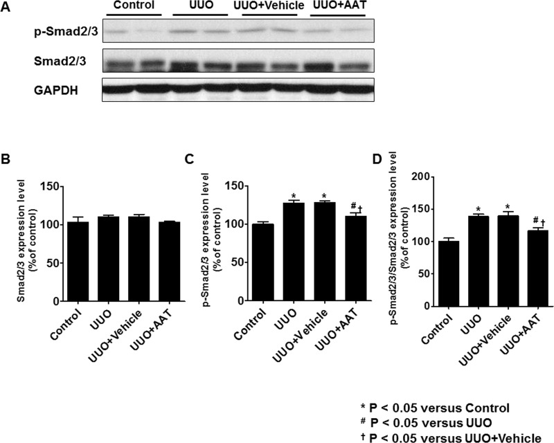
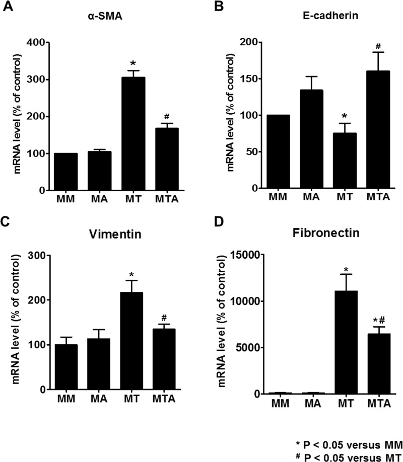
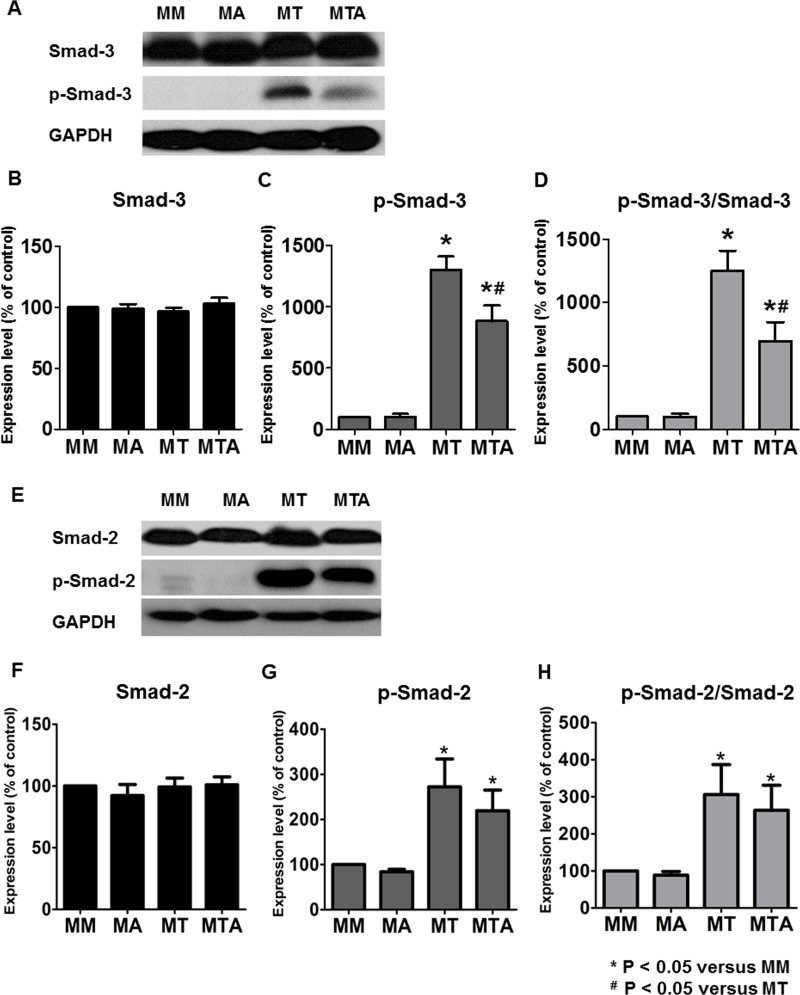
Alpha1-antitrypsin (AAT) exerts its anti-inflammatory effect through regulating the activity of serine proteinases. This study evaluated the inhibitory effects of AAT against the transforming growth factor (TGF)-β1 induced epithelial-to-mesenchymal transition (EMT) in unilateral ureter obstruction (UUO) mice and Madin-Darby canine kidney (MDCK) cells. C57BL/6 mice with induced UUO were injected intraperitoneally with AAT (80 mg/Kg) or vehicle for 7 days. MDCK cells were treated with TGF-β1 (2 ng/mL) for 48 hours to induce EMT, and co-treated with AAT (10 mg/mL) to inhibit the EMT. Masson's trichrome and Sirius red staining was used to estimate the extent of renal fibrosis in UUO mice. The expression of alpha-smooth muscle actin (α-SMA), vimentin, fibronectin, collagen I, and E-cadherin in MDCK cells and kidney tissue were evaluated. Masson's and Sirius red staining revealed that the area of renal fibrosis was significantly smaller in AAT treated UUO group compared with that of UUO and vehicle treated UUO groups. AAT treatment attenuated upregulation of Smad2/3 phosphorylation in UUO mouse model. Co-treatment of MDCK cells with TGF-β1 and AAT significantly attenuated the changes in the expression of α-SMA, vimentin, fibronectin, collagen I, and E-cadherin. AAT also decreased the phosphorylated Smad3 expression and the phosphorylated Smad3/Smad3 ratio in MDCK cells. AAT treatment inhibited EMT induced by TGF-β1 in MDCK cells and attenuated renal fibrosis in the UUO mouse model. The results of this work suggest that AAT could inhibit the process of EMT through the suppression of TGF-β/Smad3 signaling.


Conflict of interest statement

The authors have declared that no competing interests exist.

Figures

Fig 1
Fig 1. Masson’s trichrome and Sirius red staining for assessing renal fibrosis area at day 7 after unilateral ureteral obstruction (UUO).
(A and E) Sham-operation (Control). (B and F) UUO. (C and G) UUO treated with 9% NaCl (UUO + Vehicle). (D and H) UUO treated with 80 mg/Kg AAT (UUO + AAT). (I) Semi-quantitative assessment of renal fibrosis area.
Fig 2
Fig 2. Expression of α-SMA, E-cadherin, fibronectin and collagen I in the kidneys of mice with UUO.
Immunoperoxidase microscopy showed increased labeling intensity of α-SMA, fibronectin, and collagen I at the interstitial area in the UUO (B, J, and N) and vehicle treated UUO (C, K, and O) kidneys compared to that in the sham operated control kidneys (A, I, and M). In contrast, UUO (F) and vehicle treated UUO (G) kidneys showed decreased labeling intensity of E-cadherin compared to that in the control kidneys (E). AAT treatment reversed the changes in the labeling intensities of α-SMA, E-cadherin, fibronectin, and collagen I in the UUO kidneys (D, H, L, and P).
Fig 3
Fig 3. Expression of α-SMA, E-cadherin, fibronectin, and collagen I in UUO kidneys.
Expression of α-SMA, fibronectin, and collagen I were upregulated in UUO and vehicle treated UUO kidneys. Treatment with AAT significantly decreased the change in the expression of α-SMA, fibronectin, and collagen I of UUO kidneys. In contrast, the protein level of E-cadherin decreased in UUO and vehicle treated UUO kidneys and increased significantly by AAT treatment.
Fig 4
Fig 4. The mRNA level of α-SMA, E-cadherin, and collagen I in kidney tissue.
Real time RT-PCR analysis revealed significant increase in the expression of α-SMA and collagen I mRNA and decrease in E-cadherin mRNA expression in the UUO group compared to that in the control group. AAT treatment (UUO + AAT) significantly inhibited the upregulation of collagen I and downregulation of E-cadherin mRNA level compared to that in UUO alone or UUO + Vehicle treatments. AAT treatment also showed a tendency to inhibit the upregulation of α-SMA mRNA levels in the UUO kidneys.
Fig 5
Fig 5. Expression of Smad2/3 and phosphorylated Smad2/3 (p-Smad2/3) in UUO kidneys.
The level of p-Smad2/3 and the ratio of p-Smad2/3 to Smad2/3 were significantly increased in the UUO and vehicle treated UUO groups. After treatment with AAT, UUO kidneys showed significant decrease in the level of p-Smad2/3 and the ratio of p-Smad2/3 to Smad2/3 compared to UUO kidneys.
Fig 6
Fig 6. Expression of EMT markers in MDCK cells treated with TGF-β1 and AAT.
TGF-β1 exposure increased the expression of α-SMA (A and B) and decreased the expression of E-cadherin (A and C), which were reversed by AAT co-treatment. Vimentin, fibronectin, and collagen I significantly increased after TGF-β1 treatment and decreased after co-treatment with TGF-β1 and AAT (D to G). The data were normalized relative to GAPDH control. MM, cells were cultured in medium for 24 hours and then in fresh medium for additional 48 hours; MA, cells were cultured in medium for 24 hours and then in medium containing AAT for additional 48 hours; MT, cells were cultured in medium for 24 hours and then in medium containing TGF-β1 for additional 48 hours; MTA, cells were cultured in medium for 24 hours and then in medium containing TGF-β1 and AAT for additional 48 hours.
Fig 7
Fig 7. The mRNA level of α-SMA, E-cadherin, vimentin, and fibronectin in MDCK cells.
MDCK cells exposed to TGF-β1 showed upregulation of α-SMA, vimentin, and fibronectin mRNA and downregulation of E-cadherin mRNA. Co-treatment with AAT and TGF-β1 ameliorated the changes in the expression of α-SMA, vimentin, fibronectin, and E-cadherin mRNA levels in MDCK cells.
Fig 8
Fig 8
Immunofluorescence microscopy of α-SMA (A to D) and E-cadherin (E to H) in MDCK cells after TGF-β1 and AAT treatment. AAT did not cause significant change in MDCK cells cultured in medium alone (B and F). The increased immunolabeling intensity of α-SMA (C, red) seen in response to TGF-β1 treatment was attenuated by AAT treatment (D). The decreased immunolabeling intensity of E-cadherin (G, green) in response to TGF-β1 treatment was augmented by AAT treatment (H). The blue staining (A to H) is nuclear counterstaining with DAPI.
Fig 9
Fig 9. Expression of Smad3 and Smad2 in MDCK cells.
Immunoblotting showed that the phosphorylated Smad3 (p-Smad3) level and the ratio of p-Smad3 to Smad3 were significantly decreased in the MDCK cells treated with TGF-β1 and AAT compared to that in MDCK cells treated with TGF-β1 alone (A to D). The level of phosphorylated Smad2 (p-Smad2) and the ratio of p-Smad2 to Smad2 were significantly increased in MDCK cells after treatment with TGF-β1 and showed tendency to decrease in MDCK cells treated with TGF-β1 and AAT (E to H).

Similar articles

Cited by

References

    1. Liu Y (2011) Cellular and molecular mechanisms of renal fibrosis. Nat Rev Nephrol 7: 684–696. 10.1038/nrneph.2011.149 - DOI - PMC - PubMed
    1. Iwano M, Plieth D, Danoff TM, Xue C, Okada H, et al. (2002) Evidence that fibroblasts derive from epithelium during tissue fibrosis. J Clin Invest 110: 341–350. - PMC - PubMed
    1. Liu Y (2010) New insights into epithelial-mesenchymal transition in kidney fibrosis. J Am Soc Nephrol 21: 212–222. 10.1681/ASN.2008121226 - DOI - PMC - PubMed
    1. Barnes JL, Glass WF (2011) Renal interstitial fibrosis: a critical evaluation of the origin of myofibroblasts. Contrib Nephrol 169: 73–93. 10.1159/000313946 - DOI - PubMed
    1. Kriz W, Kaissling B, Le Hir M (2011) Epithelial-mesenchymal transition (EMT) in kidney fibrosis: fact or fantasy? J Clin Invest 121: 468–474. - PMC - PubMed

MeSH terms