. 2016 Sep 8;7(9):e2360.
doi: 10.1038/cddis.2016.265.

Omentin protects against LPS-induced ARDS through suppressing pulmonary inflammation and promoting endothelial barrier via an Akt/eNOS-dependent mechanism

Affiliations

Omentin protects against LPS-induced ARDS through suppressing pulmonary inflammation and promoting endothelial barrier via an Akt/eNOS-dependent mechanism

Di Qi et al. Cell Death Dis. .

Abstract

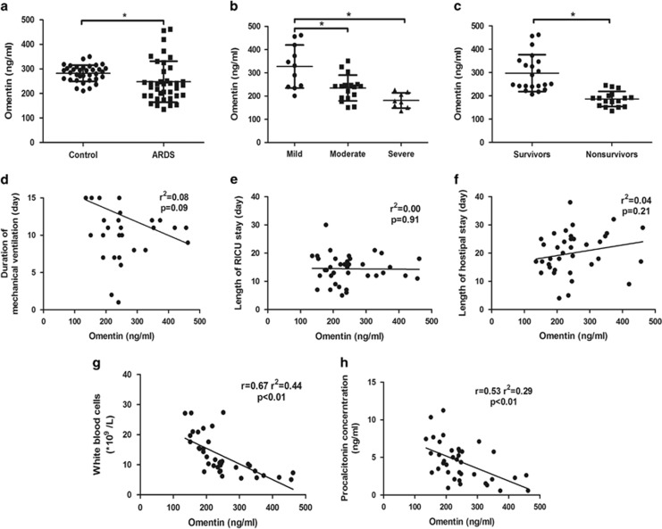
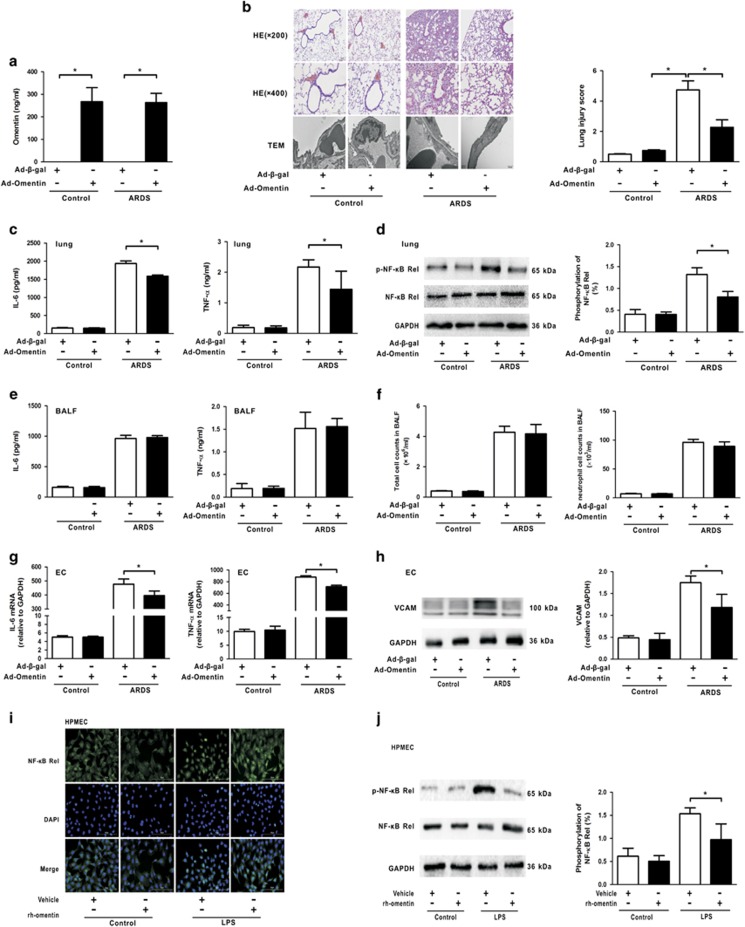
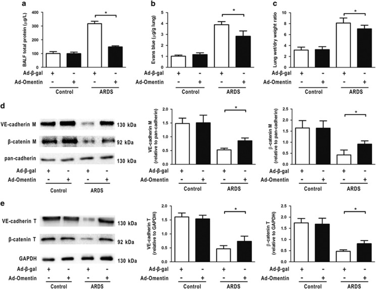
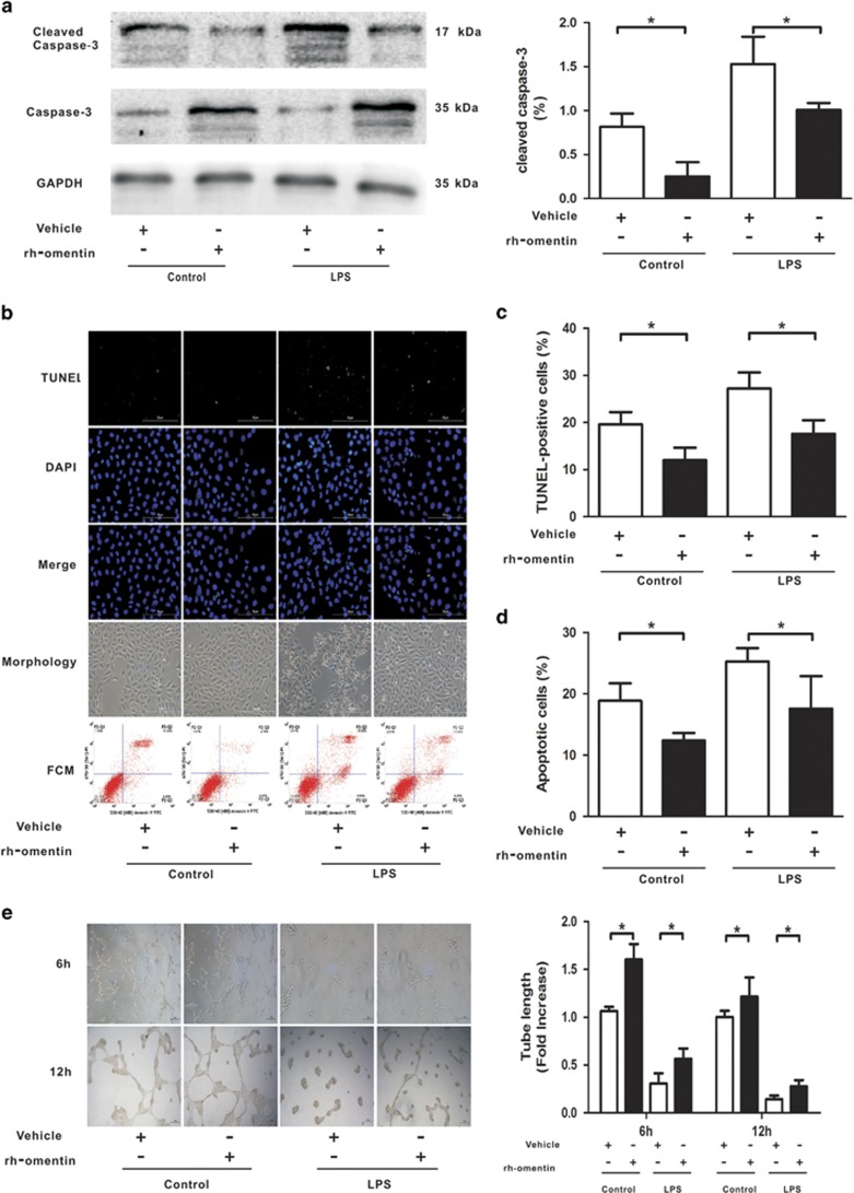
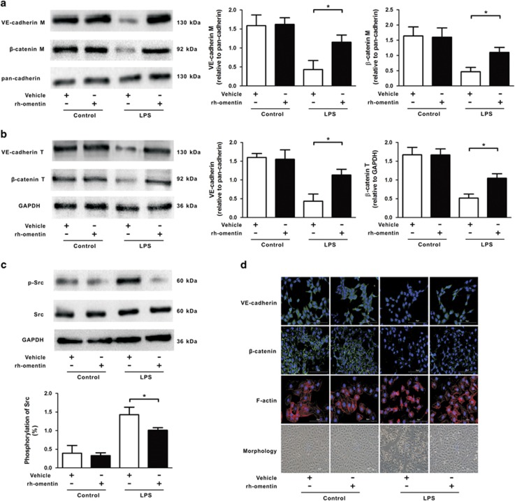
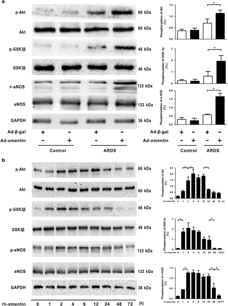
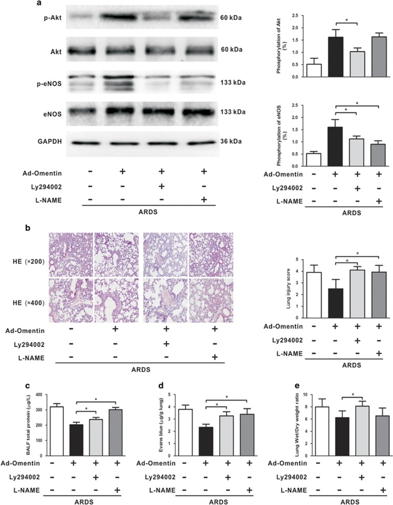
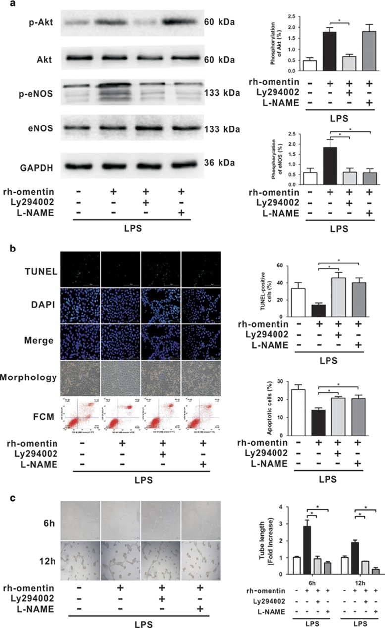
Acute respiratory distress syndrome (ARDS) is characterized by increased pulmonary inflammation and endothelial barrier permeability. Omentin has been shown to benefit obesity-related systemic vascular diseases; however, its effects on ARDS are unknown. In the present study, the level of circulating omentin in patients with ARDS was assessed to appraise its clinical significance in ARDS. Mice were subjected to systemic administration of adenoviral vector expressing omentin (Ad-omentin) and one-shot treatment of recombinant human omentin (rh-omentin) to examine omentin's effects on lipopolysaccharide (LPS)-induced ARDS. Pulmonary endothelial cells (ECs) were treated with rh-omentin to further investigate its underlying mechanism. We found that a decreased level of circulating omentin negatively correlated with white blood cells and procalcitonin in patients with ARDS. Ad-omentin protected against LPS-induced ARDS by alleviating the pulmonary inflammatory response and endothelial barrier injury in mice, accompanied by Akt/eNOS pathway activation. Treatment of pulmonary ECs with rh-omentin attenuated inflammatory response and restored adherens junctions (AJs), and cytoskeleton organization promoted endothelial barrier after LPS insult. Moreover, the omentin-mediated enhancement of EC survival and differentiation was blocked by the Akt/eNOS pathway inactivation. Therapeutic rh-omentin treatment also effectively protected against LPS-induced ARDS via the Akt/eNOS pathway. Collectively, these data indicated that omentin protects against LPS-induced ARDS by suppressing inflammation and promoting the pulmonary endothelial barrier, at least partially, through an Akt/eNOS-dependent mechanism. Therapeutic strategies aiming to restore omentin levels may be valuable for the prevention or treatment of ARDS.


Figures

Figure 1
Figure 1
Circulating omentin levels are associated with the inflammation in patients with acute respiratory distress syndrome (ARDS). (a) Plasma omentin concentrations were lower in patients with ARDS (n=38) than in healthy controls (n=35). (b) Higher omentin levels were observed in mild ARDS patients. (c) Higher omentin levels were maintained in survivors. The omentin levels were not correlated with the duration of ventilation (d), the length of RICU stay (e) and the length of hospital stay (f) in patients with ARDS. The omentin levels negatively correlated with white blood cells (g) and procalcitonin (h) in patients with ARDS. The data are presented as the mean±S.D. The lines indicate the mean value and the error bars indicate the S.D. *P<0.05
Figure 2
Figure 2
Omentin suppresses pulmonary inflammation in mouse lung tissue and pulmonary endothelial cells (ECs). Ad-omentin or Ad-β-gal as control (3 × 10⁷ PFU total per mouse) was injected into the internal jugular vein of mice for 3 days. (a) On the third day following adenovirous injection, the circulating omentin levels in Ad-omentin-treated mice increased to 267.8±42.7 ng/ml. Mice were subjected to intratracheal injection with LPS (5 mg/kg) or PBS. At 4 h after LPS injection, lung lobes were isolated to evaluate the histology and ultrastructural pathological damages. (b) H&E staining (magnification, × 200 and × 400) demonstrated that Ad-omentin significantly attenuated LPS-induced lung histopathologic alterations, including neutrophil infiltration, perivascular exudates, thickened alveolar septum, intra-alveolar and interstitial edema fluid, and hemorrhage. Lung injury scores were utilized for the quantitative analysis of lung histopathologic damage. Transmission electron microscopy showed that LPS-induced ultrastructural injuries in pulmonary ECs were significantly alleviated by Ad-omentin (n=6 independent mice from each group assayed in triplicate). At 4 h after LPS instillation, lung tissue and bronchoalveolar lavage fluid (BALF) were collected. (c) In lung tissue, Ad-omentin diminished the levels of IL-6 and TNF-α after LPS instillation (n=6 independent mice from each group assayed in triplicate). (d) In lung tissue, Ad-omentin inhibited the phosphorylation of the NF-κB Rel subunit after LPS instillation (n=6 independent mice from each group analyzed in triplicate). (e) In BALF, there were no significant differences in the levels of IL-6 and TNF-α (n=6 independent mice from each group analyzed in triplicate). (f) In BALF, there were no significant differences in the total cell and neutrophil counts (n=6 independent mice from each group analyzed in triplicate). (g) Quanitative RT-PCR analysis showed the gene levels of TNF-α and IL-6, normalized to the mRNA expression of GAPDH, were reduced in pulmonary ECs isolated from Ad-omentin-pretreated mice after LPS instillation (n=6 independent mice from each group analyzed in triplicate). (h) Western blot analysis showed that the protein levels of VCAM were decreased in pulmonary ECs isolated from Ad-omentin-pretreated mice after LPS instillation (n=6 independent mice from each group analyzed in triplicate). The relative abundances of protein bands were quantified by measuring the corresponding band intensities; the relative values are expressed normalized to GAPDH signals as shown in the bar graphs. (i) Administration of rh-omentin diminished the nuclear translocation of the NF-κB Rel subunit in HPMECs 2 h after LPS insult (n=3 independent cultures from each group assayed in triplicate, magnification, × 400). (j) Western blot analysis showed that the administration of rh-omentin reduced the phosphorylation of the NF-κB Rel subunit in HPMECs 2 h after LPS insult (n=3 independent cultures from each group assayed in triplicate). The relative abundances of protein bands were quantified by measuring the corresponding band intensities; the phosphorylation levels of NF-κB Rel subunit are expressed normalized to the total NF-κB Rel subunit signals as shown in the bar graphs. The data are presented as the mean±S.D. *P<0.05
Figure 3
Figure 3
Omentin promotes pulmonary endothelial barrier function after LPS insult in mice. Mice were systemically pre-treated with Ad-omentin or Ad-β-gal as a control (3 × 10⁷ PFU total per mouse) and subjected to intratracheal injection with LPS (5 mg/kg) or PBS. Ad-omentin significantly reduced the total BALF protein concentrations (a), EBDA extravasation (b) and W/D ratios (c) in a murine model of ARDS (n=6 independent mice from each group analyzed in triplicate). (d) Western blot analysis showed that Ad-omentin significantly reversed LPS-induced reduction in the membrance protein expression of VE-cadherin and β-catenin in mouse lung tissue after LPS instillation (n=6 independent mice from each group analyzed in triplicate). The relative abundances of protein bands were quantified by measuring the corresponding band intensities; the relative values are expressed normalized to pan-cadherin signals as shown in the bar graphs. (e) Western blot analysis showed that Ad-omentin significantly reversed LPS-induced reduction in the total protein expression of VE-cadherin and β-catenin in mouse lung tissue after LPS instillation (n=6 independent mice from each group analyzed in triplicate). The relative abundances of protein bands were quantified by measuring the corresponding band intensities; the relative values are expressed normalized to GAPDH signals as shown in the bar graphs.The data are presented as the mean±S.D. *P<0.05
Figure 4
Figure 4
Omentin improves pulmonary EC survival and differentiation after LPS insult in HPMECs. HPMECs were cultured with rh-omentin (300 ng/ml) or PBS for 24 h and exposed to either PBS or LPS (100 ng/ml) for 2 h. (a) Western blot analysis showed that rh-omentin reduced the level of cleaved caspase-3 in cultured HPMECs under both unstressed and LPS insult conditions (n=5 independent cultures from each group assayed in triplicate). The relative abundances of protein bands were quantified by measuring the corresponding band intensities; the relative values are expressed normalized to GAPDH signals as shown in the bar graphs. (b) TdT-mediated dUTP nick end labeling (TUNEL) staining and flow cytometry showed that rh-omentin diminished the ratios of TUNEL-positive cells and apoptotic cells under both unstressed and LPS insult conditions (n=3 independent cultures from each group assayed in triplicate, magnification, × 400). (c) Quantitative analyzes of TUNEL-positive HPMECs are shown in the bar graphs. (d) Quantitative analyzes of the ratios of apoptotic cells are shown in the bar graphs. (e) Endothelial cell tube formation assay demonstrated that rh-omentin promoted EC differentiation under LPS insult conditions, as evident by the increased levels of tube length (n=3 independent cultures from each group assayed in triplicate). HPMECs were grown on 48-well plates pre-coated with Matrigel (BD, NJ, USA) at a concentration of 4 × 10⁴ cells per 200 μl. After cell pellets were added to the EGM of corresponding interventions, they were cultured in an incubator at 37 °C with 5% CO2 for 12 h and then examined under a phase-contrast microscope (BX51 Olympus, Japan) with a × 10 objective. Representative photomicrographs on the left indicate HPMECs differatation into tubes. Quantitative analyzes of tube length are shown in the bar graphs.The data are presented as the mean±S.D. *P<0.05
Figure 5
Figure 5
Omentin stabilizes pulmonary EC AJs and actin cytoskeleton after LPS insult in HPMECs. HPMECs were cultured with rh-omentin (300 ng/ml) or PBS for 24 h and exposed to either PBS or LPS (100 ng/ml) for 2 h. (a) Western blot analysis showed that rh-omentin enhanced the membrane protein expression of VE-cadherin and β-catenin under LPS insult conditions (n=5 independent cultures from each group assayed in triplicate). The relative abundances of protein bands were quantified by measuring the corresponding band intensities; the relative values are expressed normalized to pan-cadherin signals as shown in the bar graphs. (b) Western blot analysis showed that rh-omentin enhanced the total protein expression of VE-cadherin and β-catenin under LPS insult conditions (n=5 independent cultures from each group assayed in triplicate). The relative abundances of protein bands were quantified by measuring the corresponding band intensities; the relative values are expressed normalized to GAPDH signals as shown in the bar graphs. (c) Western blot analysis showed that rh-omentin diminished the level of phosphorylated Scr under LPS insult conditions (n=5 independent cultures from each group assayed in triplicate). The relative abundances of protein bands were quantified by measuring the corresponding band intensities; the relative values of the phosphorylated Scr are expressed normalized to the total Scr signals, as shown in the bar graphs. (d) Immunofluorescence staining showed that rh-omentin reversed the LPS-induced decrease in the expression of β-catenin and VE-cadherin. Phalloidin staining showed that rh-omentin inhibited cell retraction, F-actin reorganization and stress fiber formation, which was induced by LPS challenge. Cell morphology analyzes showed a transition from the flattened quiescent to rounded active endothelial phenotype under LPS insult conditions, which was reversed by rh-omentin (n=3 independent cultures from each group assayed in triplicate, magnification, × 400). The data are presented as the mean±S.D. *P<0.05
Figure 6
Figure 6
Omentin activates Akt-related signaling pathways in mice and in HPMECs. (a) Mice were systemically pre-treated with Ad-omentin or Ad-β-gal as a control (3 × 10⁷ PFU total per mouse) and subjected to intratracheal injection with LPS (5 mg/kg) or PBS. Western blot analysis demonstrated that Ad-omentin enhanced the phosphorylation of Akt (p-Akt), GSK-3β (p-GSK-3β) and eNOS (p-eNOS) in mouse lungs subjected to LPS (n=6 independent mice from each group analyzed in triplicate). (b) HPMECs were cultured with rh-omentin (300 ng/ml) or PBS for 24 h and exposed to either PBS or LPS (100 ng/ml) for 2 h. Western blot analysis showed the time-dependent changes in p-Akt, p-GSK-3β and p-eNOS in HPMECs, following rh-omentin stimulation (n=5 independent cultures from each group assayed in triplicate). The relative abundances of protein bands were quantified by measuring the corresponding band intensities; the relative phosphorylation levels of protein are expressed normalized to the total protein signals, as shown in the bar graphs. The data are presented as the mean±S.D. *P<0.05
Figure 7
Figure 7
Akt/eNOS activation participates in omentin-mediated protection of the pulmonary endothelial barrier in mice. Mice were systemically pre-treated with Ad-omentin or Ad-β-gal as control (3 × 10⁷ PFU total per mouse) and subjected to intratracheally injection with LPS (5 mg/kg) or PBS. The Akt inhibitor LY294002 (40 mg/kg, dissolved in dimethyl sulfoxide (DMSO)), eNOS inhibitor L-NAME (100 mg/kg) or vehicle (10% DMSO in PBS) was intraperitoneally injected into mice 1 h before LPS or PBS instillation. (a) Western blot analysis showed that LY294002 inhibited the phosphorylation of Akt and eNOS in LPS-induced ARDS mice (n=6 independent mice from each group analyzed in triplicate). The relative abundances of protein bands were quantified by measuring the corresponding band intensities; the relative phosphorylation levels of protein are expressed normalized to the total protein signals, as shown in the bar graphs. (b) H&E staining showed that LY294002 and L-NAME aggravated histological injury in LPS-induced ARDS mice (n=6 independent mice from each group analyzed in triplicate, magnification, × 200 and × 400). Lung injury scores were utilized for quantitative analysis of lung histopathologic damage. (c) Bar graphs showed that LY294002 and L-NAME increased BALF protein levels and exacerbated EBDA extravasation in LPS-induced ARDS mice (n=6 independent mice from each group analyzed in triplicate). The data are presented as the mean±S.D. *P<0.05
Figure 8
Figure 8
Akt/eNOS activation contributes to omentin's promoting effects on the pulmonary EC barrier in HPMECs. HPMECs were pretreated with LY294002 (50 μM), L-NAME (1 mM) or vehicle (10% DMSO in PBS) 60 min before LPS and cultured with rh-omentin (300 ng/ml) or PBS for 24 h followed by exposure to LPS (100 ng/ml) for 2 h. (a) Western blot analysis showed that LY294002 inhibited rh-omentin-induced phosphorylation of Akt and eNOS, and that L-NAME blocked rh-omentin-stimulated phosphorylation of eNOS with no effects on Akt phosphorylation (n=5 independent cultures from each group assayed in triplicate). The relative abundances of protein bands were quantified by measuring the corresponding band intensities; the relative phosphorylation levels of protein are expressed normalized to the total protein signals, as shown in the bar graphs. (b) TUNEL staining, FCM and cell morphology analyzes demonstrated that LY294002 and L-NAME suppressed rh-omentin stimulated anti-apoptotic effects on pulmonary EC apoptosis as evident by elevated ratios of TUNEL-positive cells and apoptotic cells (n=3 independent cultures from each group assayed in triplicate, magnification, × 400)). (c) Endothelial cell tube formation assay demonstrated that LY294002 and L-NAME blocked rh-omentin-promoted EC differentiation under LPS insult conditions, as evidenced by decreased levels of tube length (n=3 independent cultures from each group assayed in triplicate, magnification, × 100). Representative photomicrographs on the left indicate HPMECs differatation into tubes. Quantitative analyzes of tube length are shown in the bar graphs.The data are presented as the mean±S.D. *P<0.05

Similar articles

Cited by

References

    1. Ranieri VM, Rubenfeld GD, Thompson BT, Ferguson ND, Caldwell E, Fan E et al. Acute respiratory distress syndrome: the Berlin definition. JAMA 2012; 307: 2526–2533. - PubMed
    1. Rubenfeld GD, Caldwell E, Peabody E, Weaver J, Martin DP, Neff M et al. Incidence and outcomes of acute lung injury. N Engl J Med 2005; 353: 1685–1693. - PubMed
    1. Gando S, Kameue T, Matsuda N, Sawamura A, Hayakawa M, Kato H. Systemic inflammation and disseminated intravascular coagulation in early stage of ALI and ARDS: role of neutrophil and endothelial activation. Inflammation 2004; 28: 237–244. - PubMed
    1. Muller-Redetzky HC, Suttorp N, Witzenrath M. Dynamics of pulmonary endothelial barrier function in acute inflammation: mechanisms and therapeutic perspectives. Cell Tissue Res 2014; 355: 657–673. - PMC - PubMed
    1. Mehta D, Malik AB. Signaling mechanisms regulating endothelial permeability. Physiol Rev 2006; 86: 279–367. - PubMed

MeSH terms