. 2016 Sep 8;7(9):e2363.
doi: 10.1038/cddis.2016.274.

Hemorrhagic shock primes for lung vascular endothelial cell pyroptosis: role in pulmonary inflammation following LPS

Affiliations

Hemorrhagic shock primes for lung vascular endothelial cell pyroptosis: role in pulmonary inflammation following LPS

Jie Yang et al. Cell Death Dis. .

Abstract

Hemorrhagic shock (HS) often renders patients more susceptible to lung injury by priming for an exaggerated response to a second infectious stimulus. Acute lung injury (ALI) is a major component of multiple organ dysfunction syndrome following HS and regularly serves as a major cause of patient mortality. The lung vascular endothelium is an active organ that has a central role in the development of ALI through synthesizing and releasing of a number of inflammatory mediators. Cell pyroptosis is a caspase-1-dependent regulated cell death, which features rapid plasma membrane rupture and release of proinflammatory intracellular contents. In this study, we demonstrated an important role of HS in priming for LPS-induced lung endothelial cell (EC) pyroptosis. We showed that LPS through TLR4 activates Nlrp3 (NACHT, LRR, and PYD domains containing protein 3) inflammasome in mouse lung vascular EC, and subsequently induces caspase-1 activation. However, HS induced release of high-mobility group box 1 (HMGB1), which acting through the receptor for advanced glycation end products initiates EC endocytosis of HMGB1, and subsequently triggers a cascade of molecular events, including cathepsin B release from ruptured lysosomes followed by pyroptosome formation and caspase-1 activation. These HS-induced events enhance LPS-induced EC pyroptosis. We further showed that lung vascular EC pyroptosis significantly exaggerates lung inflammation and injury. The present study explores a novel mechanism underlying HS-primed ALI and thus presents a potential therapeutic target for post-HS ALI.


Figures

Figure 1
Figure 1
HS primes for lung endothelial cell pyroptosis in response to LPS through HMGB1-RAGE signaling. (a) WT (C57BL/6) mice, TLR4−/− mice, RAGE−/− mice, and Nlrp3−/− mice were subjected to HS (HS) or sham operation (Sham) followed by LPS or saline (SAL) i.t. at 2 h after resuscitation (n=6 per group). Some WT mice received anti-HMGB1 Ab (2 mg/kg BW) by i.p. injection 30 min before HS or sham operation (n=6 per group). Lung tissue were harvested 24 h after LPS or SAL i.t. and the histological slides were stained with Cell Death Reagent-TMR (red), Alexa Fluor 488-labeled caspase-1 FLICA (green), E-selectin (white), and Hoechst (blue). Fluorescent images were obtained by confocal microscopy (original magnification × 600, higher magnification images for the selected area are shown in the respective lower right insets). Quadruple-stained cells were considered positive for pyroptotic EC. (b) The average number of pyroptotic EC of five random fields was counted for analysis. Data are presented as mean and S.E.M. *P<0.05 compared with the groups labeled with no asterisk
Figure 2
Figure 2
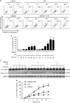
HMGB1 primes for lung EC pyroptosis in response to LPS. (ad) MLVEC isolated from WT mice were sequentially treated with HMGB1 (0.5 μg/ml) for 4 h and then with LPS (1 μg/ml) for up to 36 h. Some MLVEC were treated with HMGB1 or LPS alone. MLVEC with no treatment were as control. Cells were stained with Cell Death Reagent-TMR and Alexa Fluor 488-labeled caspase-1 FLICA, and the double-stained pyroptotic cells were detected by flow cytometry (a and b). Activation of caspase-1 in cell lysate was detected by western blot (c). IL-1β in medium was measured by ELISA (d). (eh) MLVEC from WT mice and RAGE−/− mice were sequentially treated with HMGB1 (0.5 μg/ml) for 4 h and then with LPS (1 μg/ml) for 24 h. After the treatment, cells were stained with Cell Death Reagent-TMR and Alexa Fluor 488-labeled caspase-1 FLICA, the double-stained pyroptotic cells were detected by confocal microscopy (e) and flow cytometry (f). Activation of caspase-1 in cell lysate was detected by western blot (g). IL-1β in medium was measured by ELISA (h). All images are representatives of five independent experiments, and graphs depict the value of mean and S.E.M. *P<0.05 compared with the groups labeled with no asterisk, **P<0.05 compared with the groups labeled with no or different asterisk
Figure 2
Figure 2
HMGB1 primes for lung EC pyroptosis in response to LPS. (ad) MLVEC isolated from WT mice were sequentially treated with HMGB1 (0.5 μg/ml) for 4 h and then with LPS (1 μg/ml) for up to 36 h. Some MLVEC were treated with HMGB1 or LPS alone. MLVEC with no treatment were as control. Cells were stained with Cell Death Reagent-TMR and Alexa Fluor 488-labeled caspase-1 FLICA, and the double-stained pyroptotic cells were detected by flow cytometry (a and b). Activation of caspase-1 in cell lysate was detected by western blot (c). IL-1β in medium was measured by ELISA (d). (eh) MLVEC from WT mice and RAGE−/− mice were sequentially treated with HMGB1 (0.5 μg/ml) for 4 h and then with LPS (1 μg/ml) for 24 h. After the treatment, cells were stained with Cell Death Reagent-TMR and Alexa Fluor 488-labeled caspase-1 FLICA, the double-stained pyroptotic cells were detected by confocal microscopy (e) and flow cytometry (f). Activation of caspase-1 in cell lysate was detected by western blot (g). IL-1β in medium was measured by ELISA (h). All images are representatives of five independent experiments, and graphs depict the value of mean and S.E.M. *P<0.05 compared with the groups labeled with no asterisk, **P<0.05 compared with the groups labeled with no or different asterisk
Figure 3
Figure 3
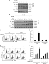
HS augments Nlrp3 inflammasome activation in lung EC through ROS-TXNIP signaling. (a) WT mice were subjected to HS (HS) or sham operation (Sham) followed by LPS (1 μg/ml) or SAL i.t. at 2 h after resuscitation. Lung tissues were recovered 12 h after LPS or SAL i.t. The association of Nlrp3-ASC was detected using immunoprecipitation with anti-ASC antibody and immunoblotting with anti-ASC and Nlrp3 antibody. The total Nlrp3 protein expression and caspase-1 p10 fragment in the lung tissue was detected by western blot. (b) WT MLVEC were sequentially treated with HMGB1 (0.5 μg/ml) for 4 h and then with LPS (1 μg/ml) for 3, 12, and 24 h. Nlrp3-ASC association, total Nlrp3, and caspase-1 p10 fragments were detected as described in (a). (cf) ROS production in live MLVEC. MLVEC from WT mice, TLR4−/− mice, and RAGE−/− mice were sequentially treated with HMGB1 (0.5 μg/ml) for 4 h and then with LPS (1 μg/ml) for 3 h. MLVEC were stained with the cell-permeable ROS detection reagent H2DFFDA (10 mM; Invitrogen Molecular Probes, Carlsbad, CA, USA) for 10 min and ROS production was then detected by flow cytometry. (g and h) WT MLVEC were sequentially treated with HMGB1 (0.5 μg/ml) for 4 h and then with LPS (1 μg/ml) for 3 h (g) and 24 h (H). For some experiments, NAC (10 mM) was added 30 min ahead of the treatment. Nlrp3 and TXNIP association was detected using immunoprecipitation and immunoblotting. IL-1β in medium was measured by ELISA. (i) TXNIP in MLVEC was knocked down using siRNA techniques as described in the Materials and Methods. At 48 h after transfection of TXNIP siRNA into MLVEC, the TXNIP protein significantly decreased as compared with control. (j and k) MLVEC were transfected with TXNIP siRNA at 48 h before treatment with HMGB1 (0.5 μg/ml) for 4 h and then with LPS (1 μg/ml) for 12 h. Nlrp3-ASC association, total Nlrp3, and caspase-1 cleavage in the EC were then detected using western blot (j) and IL-1β in the medium was measured by ELISA (k). All images are representatives of five independent experiments, and graphs depict the value of mean and S.E.M. *P<0.05 compared with the groups labeled with no or different asterisk, **P<0.05 compared with the groups labeled with no or different asterisk, #P<0.05 between the two groups
Figure 3
Figure 3
HS augments Nlrp3 inflammasome activation in lung EC through ROS-TXNIP signaling. (a) WT mice were subjected to HS (HS) or sham operation (Sham) followed by LPS (1 μg/ml) or SAL i.t. at 2 h after resuscitation. Lung tissues were recovered 12 h after LPS or SAL i.t. The association of Nlrp3-ASC was detected using immunoprecipitation with anti-ASC antibody and immunoblotting with anti-ASC and Nlrp3 antibody. The total Nlrp3 protein expression and caspase-1 p10 fragment in the lung tissue was detected by western blot. (b) WT MLVEC were sequentially treated with HMGB1 (0.5 μg/ml) for 4 h and then with LPS (1 μg/ml) for 3, 12, and 24 h. Nlrp3-ASC association, total Nlrp3, and caspase-1 p10 fragments were detected as described in (a). (cf) ROS production in live MLVEC. MLVEC from WT mice, TLR4−/− mice, and RAGE−/− mice were sequentially treated with HMGB1 (0.5 μg/ml) for 4 h and then with LPS (1 μg/ml) for 3 h. MLVEC were stained with the cell-permeable ROS detection reagent H2DFFDA (10 mM; Invitrogen Molecular Probes, Carlsbad, CA, USA) for 10 min and ROS production was then detected by flow cytometry. (g and h) WT MLVEC were sequentially treated with HMGB1 (0.5 μg/ml) for 4 h and then with LPS (1 μg/ml) for 3 h (g) and 24 h (H). For some experiments, NAC (10 mM) was added 30 min ahead of the treatment. Nlrp3 and TXNIP association was detected using immunoprecipitation and immunoblotting. IL-1β in medium was measured by ELISA. (i) TXNIP in MLVEC was knocked down using siRNA techniques as described in the Materials and Methods. At 48 h after transfection of TXNIP siRNA into MLVEC, the TXNIP protein significantly decreased as compared with control. (j and k) MLVEC were transfected with TXNIP siRNA at 48 h before treatment with HMGB1 (0.5 μg/ml) for 4 h and then with LPS (1 μg/ml) for 12 h. Nlrp3-ASC association, total Nlrp3, and caspase-1 cleavage in the EC were then detected using western blot (j) and IL-1β in the medium was measured by ELISA (k). All images are representatives of five independent experiments, and graphs depict the value of mean and S.E.M. *P<0.05 compared with the groups labeled with no or different asterisk, **P<0.05 compared with the groups labeled with no or different asterisk, #P<0.05 between the two groups
Figure 4
Figure 4
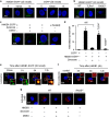
Lung EC endocytosis of HMGB1 induces lysosomal destabilization and CatB activation. (a) Confocal microscopy of MLVEC that were isolated from WT mice and incubated with recombinant HMGB1-EGFP (20 nmol/l) or EGFP for 0, 10 min, and 1 h. (b) Confocal microscopy of MLVEC that were isolated from WT, TLR4−/−, or RAGE−/− mice and incubated with HMGB1-EGFP (20 nmol/l) for 1 h. (c) Confocal microscopy of WT MLVEC that were incubated with HMGB1-EGFP in the presence or absence of dynasore (30 μg/ml) or DMSO (0.3%) for 1 h. WT MLVEC were also treated with heated HMGB1-EGFP (100 °C, 5 min) for 1 h. (d) Numerical summary of the findings from panels (a to c). Graph depicts the number of intracellular EGFP-tagged protein particles in MLVEC, which were calculated using confocal microscopy program. Mean±S.E.M, n=5. *P<0.01 compared with the groups labeled with no asterisk. (e) WT MLVEC were incubated with HMGB1-EGFP (green) and LysoTracker Red lysosome dye (red) for 30 min, 1 h, 6 h, or 12 h. Co-localization of HMGB1 and lysosome was detected using confocal microscopy. (f) MLVEC were treated with HMGB1 (0.5 μg/ml) for 3, 6, and 12 h followed by incubation with DQ ovalbumin (red) for 1 h to visualize lysosome integrity using confocal microscopy. (g) MLVEC isolated from WT or RAGE−/− mice were incubated with HMGB1 (0.5 μg/ml) for 12 h in the absence or presence of dynasore (30 μg/ml) or DMSO (0.3%). The cells were then stained with Magic Red CatB detection reagent (red) to visualize activated CatB under confocal microscopy. All images are representative of five independent experiments. Higher magnification images for the selected area are shown in the boxed insets (original magnification × 600)
Figure 5
Figure 5
HMGB1-induced CatB activation results in pyroptosome formation and caspase-1 activation. (a) Confocal microscopy of immunofluorescence of ASC foci in MLVEC. MLVEC isolated from WT mice, Nlrp3−/− mice, and RAGE−/− mice were treated with HMGB1 (0.5 μg/ml) or LPS (1 μg/ml) in the presence or absence of CA074me (10 μmol/l) for 18 h, and then ASC (red) were detected by immunofluorescence and confocal microscopy. Original magnification × 600. (b) Nlrp3−/− MLVEC were treated with HMGB1 (0.5 μg/ml) in the presence or absence of CA074me (10 μmol/l) for 6, 12, or 18 h. In one group, Nlrp3−/− MLVEC were treated with LPS alone for 18 h. Caspase-1 activation was detected by western blot. (c) WT and Nlrp3−/− MLVEC were sequentially treated with HMGB1 (0.5 μg/ml) for 4 h and then with LPS (1 μg/ml) for 3 and 18 h. IL-1β was detected by ELISA. All images are representatives of five independent experiments, and graphs depict the value of mean and S.E.M. *P<0.05 between the two groups
Figure 6
Figure 6
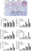
HS-primed EC pyroptosis enhances acute lung injury. (ag) WT, RAGE−/−, and caspase-1−/− mice were subjected to HS or sham operation (SM) followed by LPS or saline (SAL) i.t. at 2 h after HS. At 24 h after LPS i.t., lung histology was assessed with H&E staining (original magnification × 400) (a); lung tissue wet/dry ratio was measured (b); lung tissue MPO activity was measured using a murine MPO activity assay kit (c); protein concentration in BALF was measured by Lowry method (d); and IL-1β, IL-6, and TNF-α concentrations in BALF were determined by ELISA (eg). (hk) MLVEC isolated from WT, RAGE−/−, and caspase-1−/− were sequentially treated with HMGB1 (0.5 μg/ml) for 4 h and then with LPS (1 μg/ml) for 24 h. PMN (1 × 10⁵ cells) isolated from WT circulating blood were then added onto the surface of the treated MLVEC and incubated for 30 min, and the percentage of adherent PMN was counted under microscope (h); Endothelial permeability was assessed by Evans blue-labeled BSA (i); and IL-6, and TNF-α in the cell culture medium were measured by ELISA (j and k). (l and m) MLVEC derived from WT mice, RAGE−/− mice, and Caspase-1−/− mice were treated with HMGB1 (0.5 μg/ml) for 4 h and/or LPS (1 μg/ml) for 24 h to induce pyroptosis in the MLVEC in the upper well of Transwell, followed by co-incubating with untreated WT MLVEC, which were in the bottom well of Transwell, for additional 6 h. IL-6 and TNF-α mRNA levels in the WT MLVEC in the bottom well were then measured by qRT-PCR. All images are representatives of five independent experiments, and graphs depict the value of mean and S.E.M. *P<0.05 compared with the groups labeled with no or different asterisk, **P<0.05 compared with the groups labeled with no or different asterisk, #P<0.05 between the two groups
Figure 6
Figure 6
HS-primed EC pyroptosis enhances acute lung injury. (ag) WT, RAGE−/−, and caspase-1−/− mice were subjected to HS or sham operation (SM) followed by LPS or saline (SAL) i.t. at 2 h after HS. At 24 h after LPS i.t., lung histology was assessed with H&E staining (original magnification × 400) (a); lung tissue wet/dry ratio was measured (b); lung tissue MPO activity was measured using a murine MPO activity assay kit (c); protein concentration in BALF was measured by Lowry method (d); and IL-1β, IL-6, and TNF-α concentrations in BALF were determined by ELISA (eg). (hk) MLVEC isolated from WT, RAGE−/−, and caspase-1−/− were sequentially treated with HMGB1 (0.5 μg/ml) for 4 h and then with LPS (1 μg/ml) for 24 h. PMN (1 × 10⁵ cells) isolated from WT circulating blood were then added onto the surface of the treated MLVEC and incubated for 30 min, and the percentage of adherent PMN was counted under microscope (h); Endothelial permeability was assessed by Evans blue-labeled BSA (i); and IL-6, and TNF-α in the cell culture medium were measured by ELISA (j and k). (l and m) MLVEC derived from WT mice, RAGE−/− mice, and Caspase-1−/− mice were treated with HMGB1 (0.5 μg/ml) for 4 h and/or LPS (1 μg/ml) for 24 h to induce pyroptosis in the MLVEC in the upper well of Transwell, followed by co-incubating with untreated WT MLVEC, which were in the bottom well of Transwell, for additional 6 h. IL-6 and TNF-α mRNA levels in the WT MLVEC in the bottom well were then measured by qRT-PCR. All images are representatives of five independent experiments, and graphs depict the value of mean and S.E.M. *P<0.05 compared with the groups labeled with no or different asterisk, **P<0.05 compared with the groups labeled with no or different asterisk, #P<0.05 between the two groups
Figure 7
Figure 7
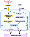
Hypothetic model for HS-primed lung EC pyroptosis and lung inflammation in response to LPS. LPS through TLR4 activates Nlrp3 inflammasome in MLVEC, and consequently induces caspase-1 activation. On the other aspect, HS induced release of HMGB1 through RAGE signaling initiates EC endocytosis of HMGB1, which in turn triggers CatB release from ruptured lysosomes followed by pyroptosome formation and caspase-1 activation. These HS-induced events enhance LPS-induced EC pyroptosis and subsequent exaggerated lung inflammation and injury

Similar articles

Cited by

References

    1. Rubenfeld GD, Caldwell E, Peabody E, Weaver J, Martin DP, Neff M et al. Incidence and outcomes of acute lung injury. N Engl J Med 2005; 353: 1685–1693. - PubMed
    1. Stapleton RD, Wang BM, Hudson LD, Rubenfeld GD, Caldwell ES, Steinberg KP. Causes and timing of death in patients with ARDS. Chest 2005; 128: 525–532. - PubMed
    1. Orfanos SE, Mavrommati I, Korovesi I, Roussos C. Pulmonary endothelium in acute lung injury: from basic science to the critically ill. Intensive Care Med 2004; 30: 1702–1714. - PubMed
    1. Matthay MA, Zimmerman GA, Esmon C, Bhattacharya J, Coller B, Doerschuk CM et al. Future research directions in acute lung injury: summary of a National Heart, Lung, and Blood Institute working group. Am J Respir Crit Care Med 2003; 167: 1027–1035. - PubMed
    1. Galluzzi L, Lopez-Soto A, Kumar S, Kroemer G. Caspases connect cell-death signaling to organismal homeostasis. Immunity 2016; 44: 221–231. - PubMed

MeSH terms

LinkOut - more resources