Clinical investigation of biofilm in non-healing wounds by high resolution microscopy techniques
- PMID: 27608736
- PMCID: PMC5058422
- DOI: 10.12968/jowc.2016.25.Sup9.S11
Clinical investigation of biofilm in non-healing wounds by high resolution microscopy techniques
Abstract
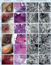
Objective: The aim of this study was to analyse wound biofilm from a clinical perspective. Research has shown that biofilm is the preferred microbial phenotype in health and disease and is present in a majority of chronic wounds. Biofilm has been linked to chronic wound inflammation, impairment in granulation tissue and epithelial migration, yet there lacks the ability to confirm the clinical presence of biofilm. This study links the clinical setting with microscopic laboratory confirmation of the presence of biofilm in carefully selected wound debridement samples.
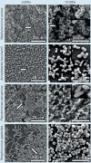
Method: Human wound debridement samples were collected from adult patients with chronic non-healing wounds who presented at the wound care centre. Sample choice was guided by an algorithm that was developed based on what is known about the characteristics of wound biofilm. The samples were then evaluated by light microscopy and scanning electron microscopy for the presence of biofilm. Details about subject history and treatment were recorded. Adherence to biofilm-based wound care (BBWC) strategies was inconsistent. Other standard antimicrobial dressings were used and no modern antiseptic wound dressings with the addition of proven antibiofilm agents were available for use.
Results: Of the patients recruited, 75% of the macroscopic samples contained biofilm despite the prior use of modern antiseptic wound dressings and in some cases, systemic antibiotics. Wounds found to contain biofilm were not all acutely infected but biofilm was present when infection was noted. The clinical histories associated with positive samples were consistent with ideas presented in the algorithm used to guide sample selection.
Conclusion: Visual cues can be used by the clinician to guide suspicion of the presence of wound biofilm. This suspicion can be further enhanced with the use of a clinical algorithm. Standard antiseptic wound dressings used in this study demonstrated limited antibiofilm efficacy. This study also highlighted a need for the clinical team to focus on expiration of dressing action and consistent practice of BBWC strategies which includes the use of proven antibiofilm agents.
Keywords: biofilm; clinical; non-healing; wound.
Figures


Similar articles
-
Biofilm in wound care.Br J Community Nurs. 2015 Mar;Suppl Wound Care:S6, S8, S10-1. doi: 10.12968/bjcn.2015.20.Sup3.S6. Br J Community Nurs. 2015. PMID: 25757387
-
[THE ROLE OF ANTISEPTICS AND STRATEGY OF BIOFILM REMOVAL IN CHRONIC WOUND].Acta Med Croatica. 2016 Mar;70(1):33-42. Acta Med Croatica. 2016. PMID: 27220188 Croatian.
-
DEVELOPMENT OF A NEXT-GENERATION ANTIMICROBIAL WOUND DRESSING.Acta Med Croatica. 2016 Mar;70(1):49-56. Acta Med Croatica. 2016. PMID: 27220190
-
Wound Biofilm: Current Perspectives and Strategies on Biofilm Disruption and Treatments.Wounds. 2017 Jun;29(6):S1-S17. Wounds. 2017. PMID: 28682297 Review.
-
Clinical experience with wound biofilm and management: a case series.Ostomy Wound Manage. 2009 Apr;55(4):38-49. Ostomy Wound Manage. 2009. PMID: 19387095 Review.
Cited by
-
Adding a new dimension: Multi-level structure and organization of mixed-species Pseudomonas aeruginosa and Staphylococcus aureus biofilms in a 4-D wound microenvironment.Biofilm. 2022 Oct 20;4:100087. doi: 10.1016/j.bioflm.2022.100087. eCollection 2022 Dec. Biofilm. 2022. PMID: 36324526 Free PMC article.
-
Clinical impact of an anti-biofilm Hydrofiber dressing in hard-to-heal wounds previously managed with traditional antimicrobial products and systemic antibiotics.Burns Trauma. 2020 Mar 4;8:tkaa004. doi: 10.1093/burnst/tkaa004. eCollection 2020. Burns Trauma. 2020. PMID: 32341917 Free PMC article.
-
Comparison Between Cultivation and Sequencing Based Approaches for Microbiota Analysis in Swabs and Biopsies of Chronic Wounds.Front Med (Lausanne). 2021 Jun 4;8:607255. doi: 10.3389/fmed.2021.607255. eCollection 2021. Front Med (Lausanne). 2021. PMID: 34150786 Free PMC article.
-
Biofilm Formation Reducing Properties of Manuka Honey and Propolis in Proteus mirabilis Rods Isolated from Chronic Wounds.Microorganisms. 2020 Nov 19;8(11):1823. doi: 10.3390/microorganisms8111823. Microorganisms. 2020. PMID: 33228072 Free PMC article.
-
A Scoping Review to Identify Clinical Signs, Symptoms and Biomarkers Reported in the Literature to Be Indicative of Biofilm in Chronic Wounds.Int Wound J. 2025 May;22(5):e70181. doi: 10.1111/iwj.70181. Int Wound J. 2025. PMID: 40389698 Free PMC article.
References
-
- Reddy M, Gill SS, Wu W, et al. Does this patient have an infection of a chronic wound? JAMA. 2012;307(6):605–611. - PubMed
-
- Seth AK, Geringer MR, Gurjala AN, et al. Treatment of Pseudomonas aeruginosa biofilm-infected wounds with clinical wound care strategies: a quantitative study using an in vivo rabbit ear model. Plast Reconstr Surg. 2012;129:262e–274e. - PubMed
-
- James GA, Swogger E, Wolcott R, et al. Biofilms in chronic wounds. Wounds Repair Regen. 2008;16:37–44. - PubMed
MeSH terms
Substances
Grants and funding
LinkOut - more resources
Full Text Sources
Other Literature Sources

