Review
. 2016 Sep 7;91(5):957-973.
doi: 10.1016/j.neuron.2016.08.027.

Lymphatics in Neurological Disorders: A Neuro-Lympho-Vascular Component of Multiple Sclerosis and Alzheimer's Disease?

Affiliations
Review

Lymphatics in Neurological Disorders: A Neuro-Lympho-Vascular Component of Multiple Sclerosis and Alzheimer's Disease?

Antoine Louveau et al. Neuron. .

Abstract

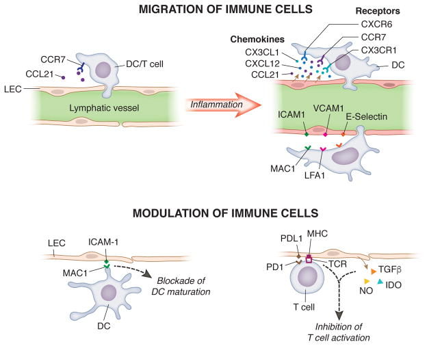
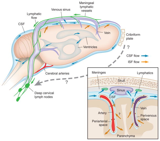
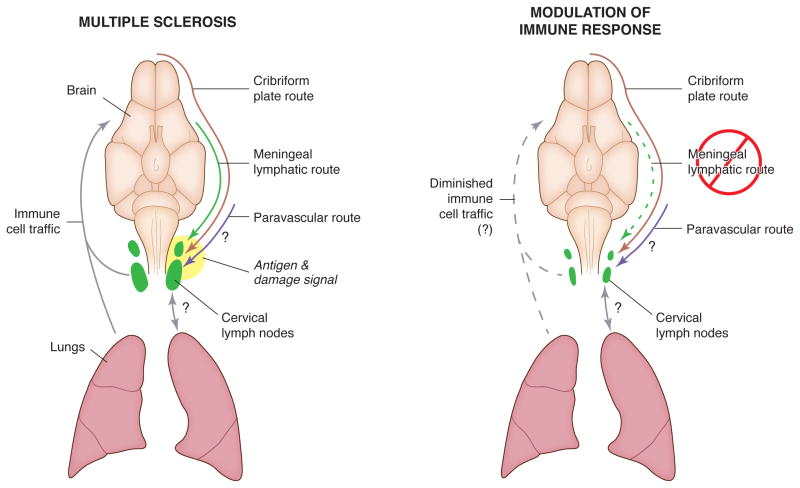
Lymphatic vasculature drains interstitial fluids, which contain the tissue's waste products, and ensures immune surveillance of the tissues, allowing immune cell recirculation. Until recently, the CNS was considered to be devoid of a conventional lymphatic vasculature. The recent discovery in the meninges of a lymphatic network that drains the CNS calls into question classic models for the drainage of macromolecules and immune cells from the CNS. In the context of neurological disorders, the presence of a lymphatic system draining the CNS potentially offers a new player and a new avenue for therapy. In this review, we will attempt to integrate the known primary functions of the tissue lymphatic vasculature that exists in peripheral organs with the proposed function of meningeal lymphatic vessels in neurological disorders, specifically multiple sclerosis and Alzheimer's disease. We propose that these (and potentially other) neurological afflictions can be viewed as diseases with a neuro-lympho-vascular component and should be therapeutically targeted as such.


Figures

Figure 1
Figure 1. Modulation of immune cell migration and function by lymphatic endothelial cells
Lymphatic endothelial cells (LECs) are able to secrete chemokine (C-C motif) ligand 21 (CCL21) and promote the entry of dendritic cells (DCs) and T cells into the lymphatic vasculature in a chemokine receptor type 7 (CCR7)-dependent manner. Upon an inflammatory stimulus, LECs produce chemokine (C-X3-C motif) ligand 1 (CX3CL1) and chemokine (C-X-C motif) ligand 12 (CXCL12), which bind the chemokine receptors CX3CR1 and CXCR4 on the surface of DCs and further facilitate their migration through the lymphatic. Adhesion and entry of the immune cells into the lymphatic vasculature is also promoted by the expression of intercellular adhesion molecule 1 (ICAM-1), vascular cell adhesion molecule 1 (VCAM-1) and E-selectin by LECs. In the lymph nodes, LECs are also able to inhibit the maturation of DCs through an ICAM-1/macrophage-1 antigen (MAC-1) dependent mechanism and the activation of T cells by releasing nitric oxide (NO), indoleamine 2,3-dioxygenase (IDO) and transforming growth factor (TGF)-β and direct interaction between major histocompatibility complex (MHC)/T cell receptor (TCR) and programmed death-ligand 1 (PD-L1)/programmed death 1 (PD-1).
Figure 2
Figure 2. Pathways of fluid circulation and drainage in the brain
The glymphatic system results from the paravascular bulk flow of interstitial fluid (ISF) into the cerebrospinal fluid (CSF) and is, at some extent, responsible for the removal of macromolecules and hydrophilic compounds from the brain parenchyma into the CSF. The circulation of ISF is driven by arterial pulsation along the basement membrane of the brain blood vessels towards the leptomeninges (orange arrows). On the other hand, the CSF recirculates from the brain ventricles to the subarachnoid meningeal space between the pia and the arachnoid mater (blue arrows). The two major routes of drainage of the aqueous content of the CSF are the arachnoid granulations, which drain directly into the major veins (sinuses) in the dura mater, and the olfactory bulbs, across the ethmoid plate, through the lymphatic network associated with the nasal mucosa. Of note, the recently described conventional meningeal lymphatic vasculature that is capable of draining macromolecules and immune cells from the meningeal spaces and the brain parenchyma into the deep cervical lymph nodes (green arrows) prompted for the reassessment of the contribution of each route to the drainage of the aqueous CSF content and, specifically, to the removal of macromolecules and immune cells from the CNS.
Figure 3
Figure 3. Hypothetical role of the meningeal lymphatic system in amyloid beta drainage from the brain fluids
The increased accumulation of amyloid beta (Aβ) peptides and consequent formation of extracellular amyloid plaques in the brain parenchyma in Alzheimer’s disease (AD) lead to the buildup of an inflammatory milieu that results in glial activation, which become inefficient in clearing extracellular amyloid, and neuronal dysfunction. The mechanisms that promote the efflux of Aβ from the brain parenchyma into the blood circulation at the neurovascular unit, as well as the production of proteins that scavenge Aβ from the CSF, become progressively obsolete with the worsening of vascular amyloid deposition (dashed arrows). Whether the function of the meningeal vasculature, and of the lymphatic vessels in particular, is also affected by the high levels of Aβ peptides present in the brain fluids in AD remains unanswered (dashed arrows). Moreover, the ability of the lymphatic vessels to modulate the levels of Aβ either by draining the subarachnoid CSF or by indirectly modulating the levels of Aβ in the ISF and its paravascular clearance through the glymphatic pathway are also important topics that warrant due attention.
Figure 4
Figure 4. Potential involvement of the meningeal lymphatic route in the CNS autoimmune response
Studies suggest that the cervical lymph nodes house macromolecules that are drained from the CNS through different pathways, namely the lymphatic network associated with the nasal mucosa (cribriform plate route, brown arrow) and the recently characterized meningeal lymphatic route (green arrow) and possibly the paravascular route (purple arrow). The cervical lymph nodes, along with the lungs, are closely involved in immune cell traffic and homing into the CNS (grey arrows) in autoimmune diseases, such as multiple sclerosis, and participate in the generation of encephalitogenic immune cells. Whether the modulation of the lymphatic drainage might attenuate antigen drainage (and associated damage signals) into the cervical lymph nodes and epitope spreading, hence decreasing the autoimmune response, and prevent the occurrence of relapses in multiple sclerosis is an important subject that needs to be addressed in the future.

References

    1. Abramov AY, Canevari L, Duchen MR. Beta-amyloid peptides induce mitochondrial dysfunction and oxidative stress in astrocytes and death of neurons through activation of NADPH oxidase. J Neurosci Off J Soc Neurosci. 2004;24:565–575. - PMC - PubMed
    1. Aebischer D, Iolyeva M, Halin C. The inflammatory response of lymphatic endothelium. Angiogenesis. 2014;17:383–393. - PubMed
    1. Akanuma S, Ohtsuki S, Doi Y, Tachikawa M, Ito S, Hori S, Asashima T, Hashimoto T, Yamada K, Ueda K, et al. ATP-binding cassette transporter A1 (ABCA1) deficiency does not attenuate the brain-to-blood efflux transport of human amyloid-beta peptide (1–40) at the blood-brain barrier. Neurochem Int. 2008;52:956–961. - PubMed
    1. D’Alessio S, Correale C, Tacconi C, Gandelli A, Pietrogrande G, Vetrano S, Genua M, Arena V, Spinelli A, Peyrin-Biroulet L, et al. VEGF-C–dependent stimulation of lymphatic function ameliorates experimental inflammatory bowel disease. J Clin Invest. 2014;124:3863–3878. - PMC - PubMed
    1. Alitalo K. The lymphatic vasculature in disease. Nat Med. 2011;17:1371–1380. - PubMed

MeSH terms