. 2016 Sep 9:6:32960.
doi: 10.1038/srep32960.

HMGB4 is expressed by neuronal cells and affects the expression of genes involved in neural differentiation

Affiliations

HMGB4 is expressed by neuronal cells and affects the expression of genes involved in neural differentiation

Ari Rouhiainen et al. Sci Rep. .

Abstract

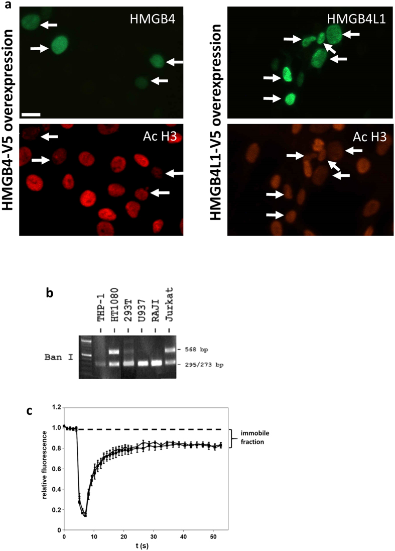
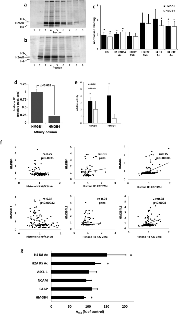
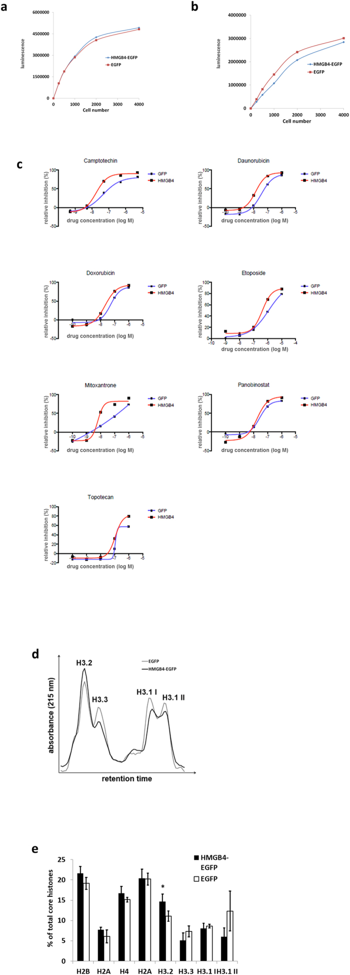
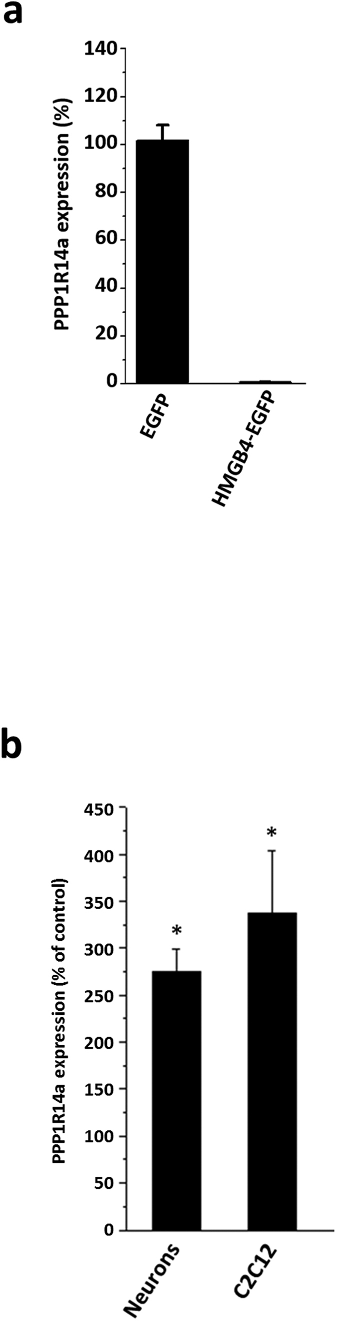
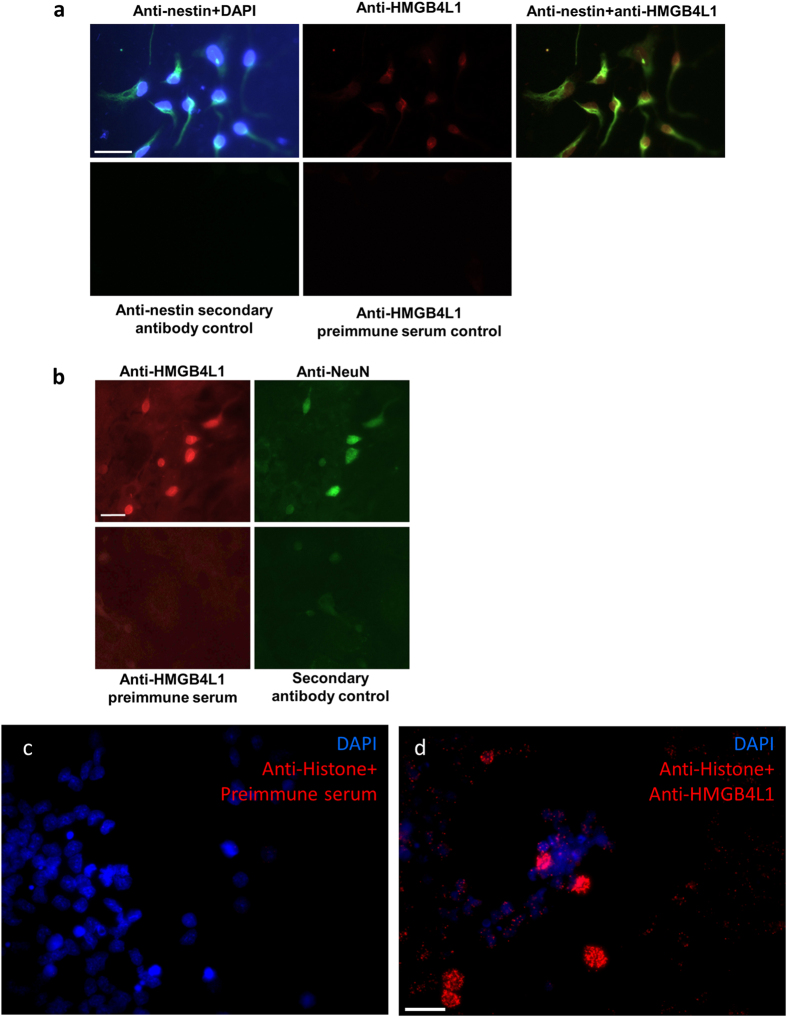
HMGB4 is a new member in the family of HMGB proteins that has been characterized in sperm cells, but little is known about its functions in somatic cells. Here we show that HMGB4 and the highly similar rat Transition Protein 4 (HMGB4L1) are expressed in neuronal cells. Both proteins had slow mobility in nucleus of living NIH-3T3 cells. They interacted with histones and their differential expression in transformed cells of the nervous system altered the post-translational modification statuses of histones in vitro. Overexpression of HMGB4 in HEK 293T cells made cells more susceptible to cell death induced by topoisomerase inhibitors in an oncology drug screening array and altered variant composition of histone H3. HMGB4 regulated over 800 genes in HEK 293T cells with a p-value ≤0.013 (n = 3) in a microarray analysis and displayed strongest association with adhesion and histone H2A -processes. In neuronal and transformed cells HMGB4 regulated the expression of an oligodendrocyte marker gene PPP1R14a and other neuronal differentiation marker genes. In conclusion, our data suggests that HMGB4 is a factor that regulates chromatin and expression of neuronal differentiation markers.


Figures

Figure 1
Figure 1. Characterization of HMGB4 and HMGB4L1.
(a) Alignment of rat TP4/HMGB4L1 and HMGB4 amino acid sequences. HMGB-boxes A and B are underlined. Red letters indicate identical amino acids. An alternative allele in position 34 of TP4/HMGB4L1 is either a tyrosine or a isoleucine. Amino acids marked in italics have been identified by amino terminal amino acid sequencing. Domain structures and number of amino acids of rat HMGB1, HMGB4 and TP4/HMGB4L1 are shown in the schematic picture. (b) Western –blot of recombinant mouse HMGB4. Recombinant HMGB4 was detected with Ponceau S –staining and with anti-HMGB4 and anti-HMGB4L1 antibodies. The antibodies did not detect recombinant HMGB1. (c) Northern Blot -analysis of the mouse HMGB4 transcript. Total RNA samples were isolated from adult mouse testes and analyzed via Northern Blot, using a probe derived from the entire coding sequence of mouse HMGB4. The probe detected a 1.1 kb band. Ethidum bromide stained ribosomal RNA is shown. (d) Northern Blot -analysis of rat HMGB4L1 transcript. Total RNA samples were isolated from adult rat testes and from developing rat testes, and analyzed via Northern Blot, using a probe derived from the entire coding sequence of rat HMGB4L1. The probe detected a 0.9 kb band in samples derived from testes of sexually mature rat. Ethidum bromide stained ribosomal RNA is shown. 3 w = 3 week old rat, 5 w = 5 week old rat. (e) Immunohistochemical staining of rat HMGB4L1 protein and in situ hybridization of mouse HMGB4 mRNA in adult testes. Anti-HMGB4L1 polyclonal antibody staining revealed intense HMGB4L1 expression in elongated spermatids (red). Haematoxylin was used as a counterstain. Control sections were stained without the primary antibody. HMGB4 mRNA localized to round and elongated spermatids. The control section shows adult mouse testes incubated with the sense probe. Bars represent 50 μm. (f) Expression of HMGB4 and HMGB4L1 mRNA in cultured rat neurons. Neuronal cells from the hippocampus and the cerebellum were cultured, and both HMGB4 and HMGB4L1 expression (arrows) was detected with RT-PCR. +RT =  reverse transcribed, -RT =  without reverse transcription.
Figure 2
Figure 2. Nuclear localization of HMGB4 and HMGB4L1.
(a) Rat glioblastoma C6 –cells transiently transfected with V5-tagged HMGB4 and HMGB4L1 were double immunostained with anti-V5-tag and anti-acetylated K9/K14 histone H3 antibodies. Immunofluorescence staining intensity of anti-acetylated K9/K14 histone H3 is reduced in C6 –cells transiently expressing HMGB4 or HMGB4L1. Arrows indicate HMGB4- and HMGB4L1-positive nuclei in double immunostained cells. Scale bar 20 = μm. (b) Single nucleotide polymorphism of the human HMGB4 gene. Genomic DNA of different human cell lines was amplified in PCR with primers specific to HMGB4 gene and subjected to Ban 1 restriction enzyme analysis. SNP rs10379 destroys Ban 1 cleavage site in the coding sequence of HMGB4 gene. Gel figure indicates that HT1080 and Jurkat cells are heterozygotes for rs10379 SNP. (c) Nuclear mobility FRAP analyses of wild type HMGB4 and rs10379 polymorphic human HMGB4 protein forms. NIH-3T3 -cells were transfected with an expression vector coding for the prominent allele or the polymorphic allele of human HMGB4-EGFP fusion proteins. One brightly fluorescent area in the nucleus with high expression of HMGB4-EGFP was bleached with three laser pulses and recovery of fluorescence was measured. Values from pre-bleach areas were determined as 1 and normalized values for bleached areas were calculated. The failing of full fluorescent recovery indicates the existence of an immobile fraction of HMGB4-EGFP in the nucleus. Triangles = wild type HMGB4, squares = SNP form of HMGB4; n = 5; ± SD.
Figure 3
Figure 3. Interactions of HMGB4 and HMGB4L1 with histones.
(a,b) Binding of C6 -cell nuclear proteins to HMGB-protein affinity columns. Proteins in elution fractions were analyzed with silver stained SDS-PAGE. The bands, representing core histones, are indicted with marks on the left. a = HMGB1 –affinity column, b = HMGB4 –affinity column. (c) Core histones eluted from HMGB-protein affinity columns and histones were quantified with ELISA. Affinity chromatography was done as described above (n ≥ 3, *p < 0.05, ±SD; Ac = acetylated, 2Me = di-methylated, 3Me = tri-methylated (lysine)). (d) HMGB1 and HMGB4 differ in their histone H1 binding capacity. Affinity chromatography was done as described above and eluted proteins were analyzed with histone H1 ELISA. Elution peak areas were quantified (n = 3, ± SD, *p < 0.002). (e) HDACs bind to HMGB1 and HMGB4. Nuclear proteins were analyzed with affinity chromatography as described above except that eluted fractions from each column were pooled to form a single fraction. Relative HDAC and sirtuin activities in elution fractions were determined (n ≥ 3 in each experiment, ±SD). (f ) Immunofluorescence intensities of modified histone antibody stained HMGB4 or HMGB4L1 overexpressing cells. Rat glioblastoma C6- cells transiently expressing HMGB4-V5 or HMGB4L1-V5 were double-immunostained with anti-acetylated (K9/K14), anti-dimethylated K27 or anti-trimethylated K27 histone H3 antibodies and with anti-V5 antibodies. The correlation blots are shown. (g) Regulation of protein levels in neuronal precursor cells by HMGB4 shRNA. Human neuronal precursor NTERA-2 cl. D1 cells stably overexpressing HMGB4 shRNA or control nonspecific shRNA were analyzed in cell ELISA. Absorbance values of HMGB4 shRNA expressing cells were normalized to values of control shRNA expressing cells (n ≥ 3, ± SD, *p < 0.03). H2A K5 Ac = histone H2A acetylated lysine 5, H4 K8 Ac = histone H4 acetylated lysine 8.
Figure 4
Figure 4. HEK 293T -cells overexpressing HMGB4-EGFP have increased sensitivity to topoisomerase inhibitors and altered histone variant composition.
(a) Growth of HEK 293T -cell clones with doxycycline induced HMGB4-EGFP or EGFP -expression. The expression of HMGB4-EGFP or EGFP in cells was induced over one week with doxycycline and viability was measured with CellTiterGlo Luminescent assay reagent via ATP quantification. (b) Growth of stable HEK 293T -cell clones, constantly expressing HMGB4-EGFP or EGFP. Cells were cultured for 72 h and viability was measured as described above. (c) Overexpression of HMGB4 increases cell sensitivity to topoisomerase inhibitors. Cells overexpressing HMGB4-EGFP were more sensitive to topoisomerease inhibitors than control cells expressing EGFP. Figure shows representative curves from two different experiments. (d) Quantification of histone H3 variants of HMGB4-EGFP or EGFP-expressing HEK 293T -cell clones. Histones were isolated from the cells and analyzed with RP-HPLC. Histone peaks were identified according to their relative retention times. Histone H3.1 eluted in two peaks (H3.1 I and H3.1 II). Curves are derived from three EGFP -control cell clone analyses and from three HMGB4-EGFP –cell clone analyses. (e) Maximal core histone peak heights of RP-HPLC (see above) were determined with the UNICORN- software. Peak height sum of core histones was determined as 100% and relative peak heights were calculated. The relative amount of histone H3.2 was elevated in the HMGB4-EGFP -expressing cells when compared to the control cells (n = 3, ±SD, *p < 0.05).
Figure 5
Figure 5. Expression of PPP1R14a is regulated by HMGB4.
(a) Inhibition of PPP1R14a expression in HEK 293T -cells constitutively expressing HMGB4-EGFP. Three independent repeats were analyzed by comparing representative EGFP expressing control and HMGB4-EGFP expressing cells. Results from representative analysis are shown. (b) Mouse E14 cortical neurons and C2C12 myoblast cells, both expressing endogenous HMGB4 mRNA, were treated with HMGB4 Vivo-Morpholino to downregulate translation of HMGB4. The expression levels of PPP1R14a were analyzed with qPCR. The levels of the PPP1R14a transcript in HMGB4 Vivo-Morpholino -treated cells were normalized to the PPP1R14a transcript levels in control Vivo-Morpholino -treated cells (*p < 0.05, ± SEM; n = 6 in C2C12 experiment; n = 18 in neuron experiment).
Figure 6
Figure 6. Expression of HMGB4 and HMGB4L1 in cultured primary cells of rat brain.
(a) Immunofluorescence staining of nestin and HMGB4L1 in 1d differentiated rat neuronal cells in vitro. Neurospheres were allowed to adhere and differentiate for 1 day and cells were immunofluorescently stained with anti-nestin and anti-HMGB4L1 antibodies. Cell nuclei were stained with DAPI. The staining controls for anti-nestin or anti-HMGB4L1 staining were done without primary antibody or with preimmune serum, respectively. Scale bar = 30 μm. (b) Immunofluorescence staining of HMGB4L1 and NeuN in 14d differentiated rat neuronal cells in vitro. Neurospheres were allowed to differentiate for 14 days and cells were immunofluorescently stained with anti-NeuN and anti-HMGB4L1 antibodies. Staining controls for anti-HMGB4L1 and anti-NeuN were done with pre-immune serum or without primary antibody, respectively. Scale bar = 20 μm. (c,d) Proximity ligation assay of neurospheres with anti-PAN-Histone and anti-HMGB4L1 antibodies. Cell nuclei of cultured neurospheres were stained with DAPI and a proximity ligation assay was performed with anti-PAN-Histone antibodies and preimmune control serum (c) or with anti-PAN-Histone antibodies and anti-HMGB4L1 antibodies (d). Scale bar = 20 μm.

Similar articles

Cited by

References

    1. Gaillard C. & Strauss F. Association of poly(CA).poly(TG) DNA fragments into four-stranded complexes bound by HMG1 and 2. Science 264, 433–436 (1994). - PubMed
    1. Catena R. et al. HMGB4, a novel member of the HMGB family, is preferentially expressed in the mouse testis and localizes to the basal pole of elongating spermatids. Biol. Reprod. 80, 358–366 (2009). - PubMed
    1. Itou J. et al. HMGB factors are required for posterior digit developmet through integrating signaling pathway activities. Dev. Dyn. 240, 1151–1162 (2011). - PMC - PubMed
    1. Zhou Q. et al. A multipotent progenitor domain guides pancreatic organogenesis. Dev. Cell. 13, 103–114 (2007). - PubMed
    1. Abraham A. B. et al.Members of the high mobility group B protein family are dynamically expressed in embryonic neural stem cells. Proteome Sci. 11, 18 (2013). - PMC - PubMed

Publication types

LinkOut - more resources