. 2016 Sep 8;16(1):723.
doi: 10.1186/s12885-016-2742-y.

In vivo bioluminescence imaging for leptomeningeal dissemination of medulloblastoma in mouse models

Affiliations

In vivo bioluminescence imaging for leptomeningeal dissemination of medulloblastoma in mouse models

Seung Ah Choi et al. BMC Cancer. .

Abstract

Background: The primary cause of treatment failure in medulloblastomas (MB) is the development of leptomeningeal dissemination (seeding). For translational research on MB seeding, one of the major challenges is the development of reliable experimental models that simulate the seeding and growth characteristics of MBs. To overcome this obstacle, we improved an experimental mouse model by intracisternal inoculation of human MB cells and monitoring with in vivo live images.

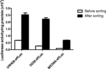
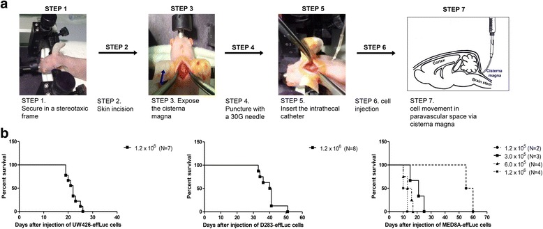
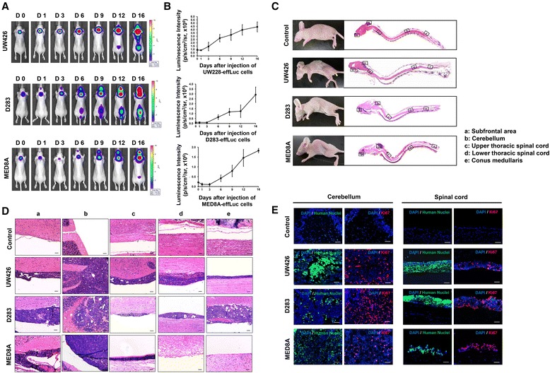
Methods: Human MB cells (UW426, D283 and MED8A) were transfected with a firefly luciferase gene and a Thy1.1 (CD90.1) marker linked with IRES under the control of the CMV promoter in a retroviral DNA backbone (effLuc). The MB-effLuc cells were injected into the cisterna magna using an intrathecal catheter, and bioluminescence images were captured. We performed histopathological analysis to confirm the extent of tumor seeding.

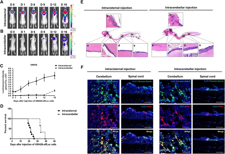
Results: The luciferase activity of MB-effLuc cells displayed a gradually increasing pattern, which correlated with a quantitative luminometric assay. Live imaging showed that the MB-effLuc cells were diffusely distributed in the cervical spinal cord and the lumbosacral area. All mice injected with UW426-effLuc, D283-effLuc and MED8A-effLuc died within 51 days. The median survival was 22, 41 and 12 days after injection of 1.2 × 10(6) UW426-effLuc, D283-effLuc and MED8A-effLuc cells, respectively. The histopathological studies revealed that the MB-effLuc cells spread extensively and diffusely along the leptomeninges of the brain and spinal cord, forming tumor cell-coated layers. The tumor cells in the subarachnoid space expressed a human nuclei marker and Ki-67. Compared with the intracerebellar injection method in which the subfrontal area and distal spinal cord were spared by tumor cell seeding in some mice, the intracisternal injection model more closely resembled the widespread leptomeningeal seeding observed in MB patients.

Conclusion: The results and described method are valuable resources for further translational research to overcome MB seeding.

Keywords: In vivo bioluminescence imaging; Intracisternal injection; Leptomeningeal seeding; Medulloblastoma.


Figures

Fig. 1
Fig. 1
Retroviral construct that contains the effLuc gene and Thy1.1 (CD90.1), linked with an internal ribosomal entry site (IRES). The luciferase activity of effLuc cells cultured in a 96-well plate was measured using an IVIS-100 optical imaging device. Firefly luciferase activity continuously increased in effLuc cells in proportion to cell number
Fig. 2
Fig. 2
Intracisternal injection for medulloblastoma (MB) seeding model and survival analysis. a Immune-deficient BALB/c-nude mice were mounted on the stereotactic device. The mouse heads were fixed in a stereotactic device, and the cisterna magna was exposed. MB-effLuc cells (UW426-effLuc: 1.2 × 106; MED8A-effLuc: 1.2 × 105, 3.0 × 105, 6.0 × 105 and 1.2 × 106; D283-effLuc: 1.2 × 106) were slowly injected into the subarachnoid space of the cisterna magna. b The median survival days of each group were estimated: UW426-effLuc: 22 days for 1.2 × 106 cells; D283-effLuc: 41 days for 1.2 × 106 cells; MED8A-effLuc: 57 days for 1.2 × 105 cells, 21 days for 3.0 × 105 cells, 14 days for 6.0 × 105 cells and 12 days for 1.2 × 106 cells
Fig. 3
Fig. 3
Bioluminescence imaging (BLI) and histological analysis for the evaluation of medulloblastoma (MB) seeding. (A and B) After injection of the MB-effLuc cells (UW426, 1.2 × 106; D283, 1.2 × 106; MED8A, 3.0 × 105), firefly luciferase BLI was evaluated and quantified at the indicated days. (C) Representative longitudinal sections of the brains and spinal cords of the mice with saline (control) or MB-effLuc cells (hematoxylin & eosin stain) (1.25×). (D) Inlet figures denote the subfrontal area (a), cerebellum (b), upper thoracic spinal cord (c), lower thoracic spinal cord (d), and conus medullaris (e). Scale bars represent 50 μm. (E) Representative immunofluorescence images (DAPI: blue, Human Nuclei: green, Ki67: red) show that the MB-effLuc cells are highly proliferative in vivo. Scale bars represent 50 μm
Fig. 4
Fig. 4
The intracisternal injection and intracerebellar injection methods. (A) BLI of mice with cell injection into the cisterna magna show that the signals are observed at day 0, expand at day 6, and begin to spread to the spinal cord at day 9. (B) The signals were detected first at day 6 and gradually migrated to the spinal cord from day 12 in mice with cells injected into the cerebellum. (C) BLI quantification of tumor-occupied areas during the study. (D) The median survival was 22 and 34 days in the intracisternal injection model and intracerebellar injection model, respectively. (E) Histopathology of xenograft MB seeding. Inlet figures denote the subfrontal area (a), cerebellum (b), upper thoracic spinal cord (c), lower thoracic spinal cord (d), and conus medullaris (e). The intracisternal injection model displays strong similarity to the histopathological character and widespread dissemination pattern of MB seeding. (F) Representative immunofluorescence images (DAPI: blue, Human Nuclei: green, Ki-67: red, Merge: yellow) show that both the intracisternal- and intracerebellar-injected UW426-effLuc cells are highly proliferative in vivo. Scale bars represent 50 μm

Similar articles

Cited by

References

    1. Phi JH, Lee J, Wang KC, Cho BK, Kim IO, Park CK, et al. Cerebrospinal fluid M staging for medulloblastoma: reappraisal of Chang’s M staging based on the CSF flow. Neuro-Oncology. 2011;13(3):334–44. doi: 10.1093/neuonc/noq171. - DOI - PMC - PubMed
    1. Young RJ, Khakoo Y, Yhu S, Wolden S, De Braganca KC, Gilheeney SW, et al. Extraneural metastases of medulloblastoma: desmoplastic variants may have prolonged survival. Pediatr Blood Cancer. 2015;62(4):611–5. doi: 10.1002/pbc.25354. - DOI - PubMed
    1. Jenkin D, Shabanah MA, Shail EA, Gray A, Hassounah M, Khafaga Y, et al. Prognostic factors for medulloblastoma. Int J Radiat Oncol Biol Phys. 2000;47(3):573–84. doi: 10.1016/S0360-3016(00)00431-4. - DOI - PubMed
    1. Phi JH, Choi SA, Lim SH, Lee J, Wang KC, Park SH, et al. ID3 contributes to cerebrospinal fluid seeding and poor prognosis in medulloblastoma. BMC Cancer. 2013;13:291. doi: 10.1186/1471-2407-13-291. - DOI - PMC - PubMed
    1. Goodrich LV, Milenkovic L, Higgins KM, Scott MP. Altered neural cell fates and medulloblastoma in mouse patched mutants. Science. 1997;277(5329):1109–13. doi: 10.1126/science.277.5329.1109. - DOI - PubMed

Publication types

LinkOut - more resources