

Ultra-wide field angiography in the management of Eales disease
- PMID: 27609162
- PMCID: PMC5026075
- DOI: 10.4103/0301-4738.190138
Ultra-wide field angiography in the management of Eales disease
Abstract
Aims: This study aims to evaluate the use of ultra-wide field (UWF) angiography in patients with Eales disease (ED).
Settings and design: Prospective observational case series in tertiary eye care center.
Subjects and methods: This study involved 17 patients diagnosed with ED, who underwent UWF fluorescein angiography. The angiograms were analyzed to look for additional information as compared to Early Treatment Diabetic Retinopathy Study seven standard field. The impact of this information in the management of patients was analyzed.
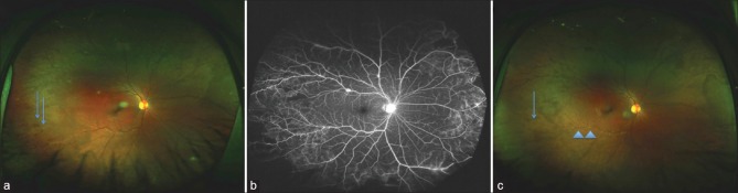
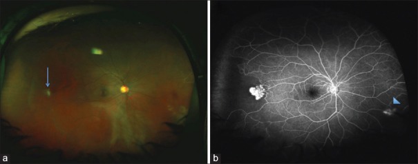
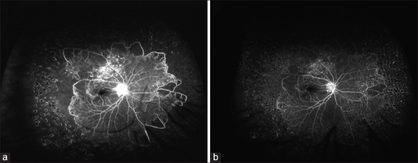
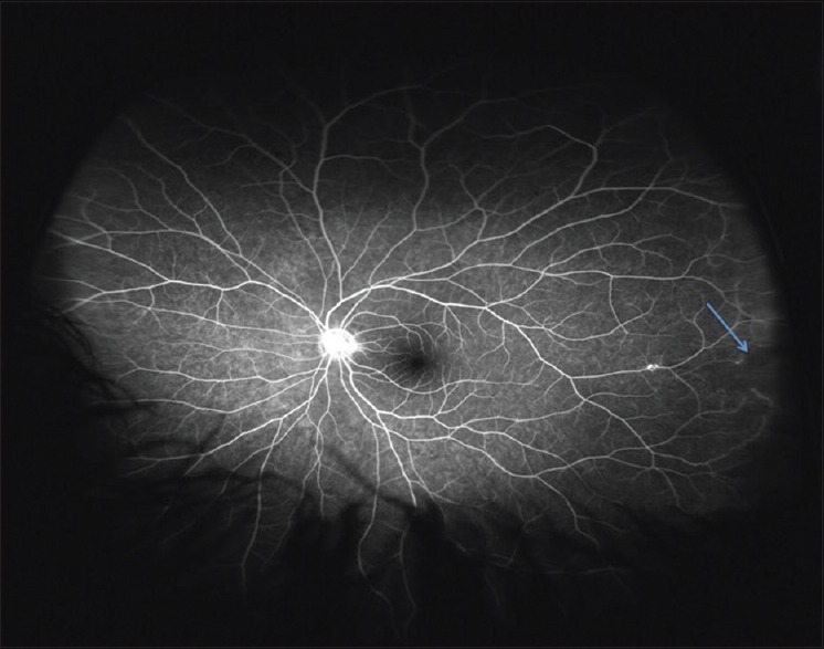
Results: 24 eyes of 17 patients with mean age of 26.3 years were diagnosed with ED and underwent UWF angiography. UWF fluorescein angiography was helpful in the documentation of peripheral retinal changes (in 67% of eyes), exact localization of capillary nonperfusion (CNP) (in 54% of eyes), and in determination of vascular involvement (in 21% of eyes). In 33% of eyes, immediate treatment plan changed because of changes picked up on UWF angiography.
Conclusions: UWF angiography helped in the better documentation, exact quantification, and location of CNP areas and better determination of disease activity. UWF imaging may play an important part in the management of patients with ED.
Figures
References
-
- Das T, Pathengay A, Hussain N, Biswas J. Eales’ disease: Diagnosis and management. Eye (Lond) 2010;24:472–82. - PubMed
-
- Puttamma ST. Varied fundus picture of central retinal vasculitis. Trans Asia Pac Acad Ophthalmol. 1970;3:520.
-
- Das T, Biswas J, Kumar A, Nagpal PN, Namperumalsamy P, Patnaik B, et al. Eales’ disease. Indian J Ophthalmol. 1994;42:3–18. - PubMed
-
- Witmer MT, Kiss S. Wide-field imaging of the retina. Surv Ophthalmol. 2013;58:143–54. - PubMed
Publication types
MeSH terms
Supplementary concepts
LinkOut - more resources
Full Text Sources
Other Literature Sources
