. 2016 Sep 12;9(17):1835-43.
doi: 10.1016/j.jcin.2016.06.020.

Intentional Laceration of the Anterior Mitral Valve Leaflet to Prevent Left Ventricular Outflow Tract Obstruction During Transcatheter Mitral Valve Replacement: Pre-Clinical Findings

Affiliations

Intentional Laceration of the Anterior Mitral Valve Leaflet to Prevent Left Ventricular Outflow Tract Obstruction During Transcatheter Mitral Valve Replacement: Pre-Clinical Findings

Jaffar M Khan et al. JACC Cardiovasc Interv. .

Abstract

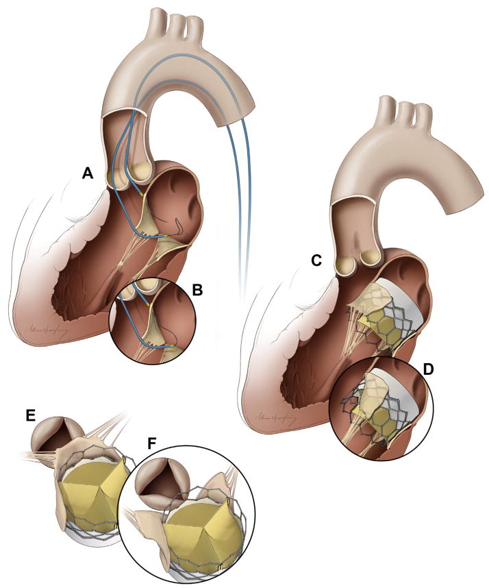
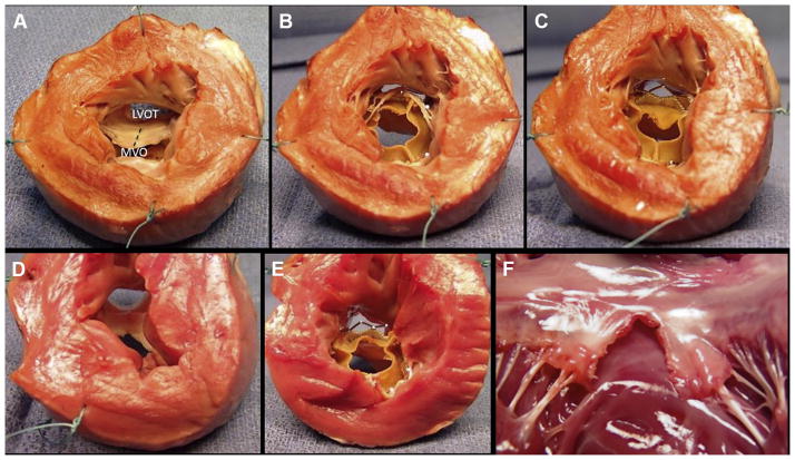
Objectives: The authors propose a novel transcatheter transection of the anterior mitral leaflet to prevent iatrogenic left ventricular outflow tract (LVOT) obstruction during transcatheter mitral valve replacement (TMVR).

Background: LVOT obstruction is a life-threatening complication of TMVR caused by septal displacement of the anterior mitral leaflet.

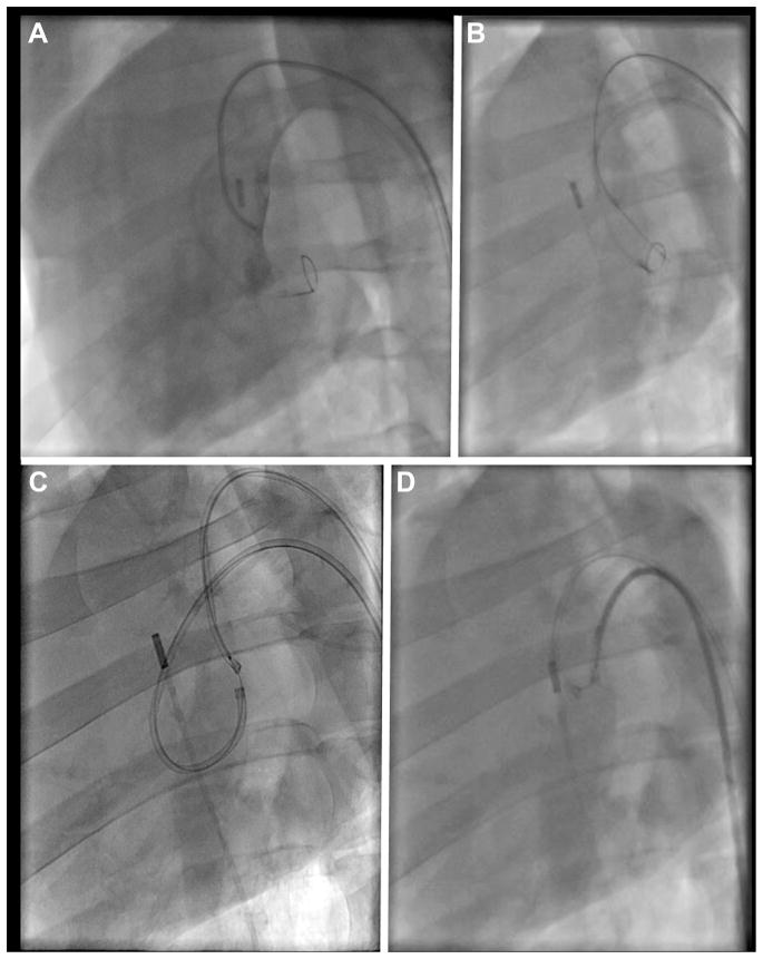
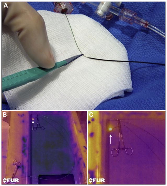
Methods: In vivo procedures in swine were guided by biplane x-ray fluoroscopy and intracardiac echocardiography. Retrograde transaortic 6-F guiding catheters straddled the anterior mitral leaflet. A stiff 0.014-inch guidewire with polymer jacket insulation was electrified and advanced from the LVOT, through the A2 leaflet base, into the left atrium. The wire was snared and externalized, forming a loop that was energized and withdrawn to lacerate the anterior mitral leaflet.

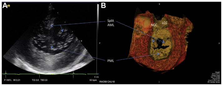
Results: The anterior mitral leaflet was successfully lacerated in 7 live and 1 post-mortem swine under heparinization. Lacerations extended to 89 ± 19% of leaflet length and were located within 0.5 ± 0.4 mm of leaflet centerline. The chordae were preserved and retracted the leaflet halves away from the LVOT. LVOT narrowing after benchtop TMVR was significantly reduced with intentional laceration of the anterior mitral leaflet to prevent LVOT obstruction than without (65 ± 10% vs. 31 ± 18% of pre-implantation diameter, p < 0.01). The technique caused mean blood pressure to fall (from 54 ± 6 mm Hg to 30 ± 4 mm Hg, p < 0.01), but blood pressure remained steady until planned euthanasia. No collateral tissue injury was identified on necropsy.

Conclusions: Using simple catheter techniques, the anterior mitral valve leaflet was transected. Cautiously applied in patients, this strategy can prevent anterior mitral leaflet displacement and LVOT obstruction caused by TMVR.

Keywords: left ventricular outflow tract obstruction; mitral valve; structural heart disease; subvalvular aortic stenosis; transcatheter mitral valve replacement; valvular heart disease.


Figures

FIGURE 1
FIGURE 1. Illustrations of the Technique of Intentional Laceration of the Anterior Mitral Valve Leaflet to Prevent Left Ventricular Outflow Tract Obstruction
(A) Two Judkins left catheters are positioned on either side of the A2 mitral leaflet base. An energized guidewire is advanced from the left ventricular outflow tract (LVOT) catheter into the left atrial catheter snare. (B) The snared tip is externalized to form a guidewire loop around the A2 leaflet. This is energized and pulled outward to lacerate the leaflet lengthwise into 2 halves. (C,E) A transcatheter mitral valve implant tents the anterior mitral leaflet into the septum, obstructing the LVOT. (D,F) Splitting the leaflet by intentional laceration of the anterior mitral valve leaflet to prevent LVOT obstruction instead causes the 2 tethered halves to displace along either side of the transcatheter valve, preventing LVOT obstruction.
FIGURE 2
FIGURE 2. Fluoroscopy Demonstration of Intentional Laceration of the Anterior Mitral Valve Leaflet to Prevent Left Ventricular Outflow Tract Obstruction in a Left Oblique Projection
(A) Angiography through the left ventricular outflow tract (LVOT) catheter shows good positioning of this catheter at the base of the anterior leaflet, below the aortic valve, with a loop snare positioned through the left atrial (LA) catheter. (B) The electrified guidewire is advanced through the A2 mitral leaflet base into the LA snare. (C) A denuded kinked section of the guidewire, insulated and marked proximally with a polymer wire convertor further insulated by the 2 guiding catheters, is electrified while the LA catheter is pulled back into the LVOT (position D) during stage 1 of the 2-step electrosurgical laceration. (D) Stage 2 of the laceration. Both catheters are pulled in tandem during a burst application of radiofrequency energy, lacerating the leaflet completely and freeing the catheter-guidewire loop.
FIGURE 3
FIGURE 3. Guidewire Electrosurgery
(A) A short midshaft section of the electrically insulating polytetrafluoroethylene coating of a 0.014-inch guidewire is stripped using a scalpel and then kinked, with a polymer jacket wire convertor locked alongside. (B) Infrared images of a saline bath with a denuded guidewire loop through 2 catheters, replicating in vivo intentional laceration of the anterior mitral valve leaflet to prevent left ventricular outflow tract obstruction. The guidewire is clipped to an electrosurgery pencil and electrified, revealing a hot spot (bright yellow, arrow) only at the exposed guidewire loop. (C) A close-up of the guidewire loop reveals no heating around the nearby metallic hemostat, suggesting freedom from electric coupling.
FIGURE 4
FIGURE 4. Images of the Lacerated Anterior Mitral Leaflet After Intentional Laceration of the Anterior Mitral Valve Leaflet to Prevent Left Ventricular Outflow Tract Obstruction
(A) Short-axis intraoperative intracardiac echocardiographic image of the mitral valve showing the anterior mitral leaflet (AML) split in 2 equal halves. (B) The corresponding post-procedural surface rendering of contrast-enhanced computed tomography also displaying split and splayed leaflets. See Online Video 1. AML = anterior mitral leaflet; LCC = left coronary cusp; NCC = noncoronary cusp; PML = posterior mitral leaflet.
FIGURE 5
FIGURE 5. Benchtop Assessment of Left Ventricular Outflow Tract Geometry Impact
The base of the left ventricle is viewed in cross section after the apex is cut away. (A) A naive heart with the anterior mitral leaflet intact. The trajectory of an intentional laceration of the anterior mitral valve leaflet to prevent left ventricular outflow tract obstruction (LAMPOON) laceration is depicted by the dashed line. (B) Transcatheter mitral valve replacement (TMVR) with intact anterior leaflet showing reduced left ventricular outflow tract (LVOT) area. (C) LAMPOON modification made to the same heart, with the anterior leaflets displaced to the side by TMVR and reduced LVOT obstruction. Flow would be possible through uncovered stent struts. (D) Explant after in vivo LAMPOON heart showing lacerated anterior leaflet. (E) TMVR in the explanted heart after LAMPOON showing displacement of the anterior leaflet away from the LVOT. (F) Explanted heart after isolated in vivo LAMPOON viewed from the posterior wall, showing central laceration down the complete length of the anterior leaflet. The intact subvalvular apparatus displaces the leaflet tips away from the LVOT. LVOT = left ventricular outflow tract; MVO = mitral valve orifice.

References

    1. Eleid MF, Cabalka AK, Williams MR, et al. Percutaneous transvenous transseptal trans-catheter valve implantation in failed bioprosthetic mitral valves, ring annuloplasty, and severe mitral annular calcification. J Am Coll Cardiol Intv. 2016;9:1161–74.
    1. Sud K, Agarwal S, Parashar A, et al. Degenerative mitral stenosis: unmet need for percutaneous interventions. Circulation. 2016;133:1594–604. - PubMed
    1. Lee KS, Stewart WJ, Lever HM, Underwood PL, Cosgrove DM. Mechanism of outflow tract obstruction causing failed mitral valve repair. Anterior displacement of leaflet coaptation. Circulation. 1993;88:II24–9. - PubMed
    1. Mihaileanu S, Marino JP, Chauvaud S, et al. Left ventricular outflow obstruction after mitral valve repair (Carpentier’s technique). Proposed mechanisms of disease. Circulation. 1988;78:I78–84. - PubMed
    1. Blanke P, Naoum C, Dvir D, et al. Predicting LVOT obstruction in transcatheter mitral valve implantation: concept of the neo-LVOT. J Am Coll Cardiol Img. In press.

Publication types

MeSH terms