

Specialized roles of neurofilament proteins in synapses: Relevance to neuropsychiatric disorders
- PMID: 27609296
- PMCID: PMC5079776
- DOI: 10.1016/j.brainresbull.2016.09.002
Specialized roles of neurofilament proteins in synapses: Relevance to neuropsychiatric disorders
Abstract
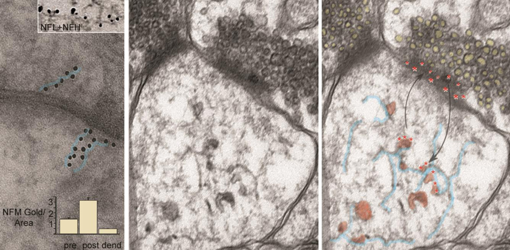
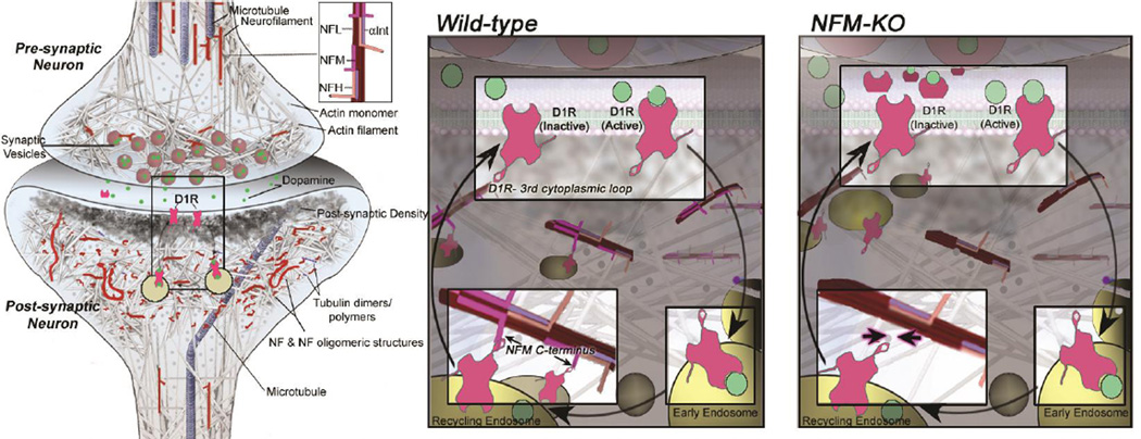
Neurofilaments are uniquely complex among classes of intermediate filaments in being composed of four subunits (NFL, NFM, NFH and alpha-internexin in the CNS) that differ in structure, regulation, and function. Although neurofilaments have been traditionally viewed as axonal structural components, recent evidence has revealed that distinctive assemblies of neurofilament subunits are integral components of synapses, especially at postsynaptic sites. Within the synaptic compartment, the individual subunits differentially modulate neurotransmission and behavior through interactions with specific neurotransmitter receptors. These newly uncovered functions suggest that alterations of neurofilament proteins not only underlie axonopathy in various neurological disorders but also may play vital roles in cognition and neuropsychiatric diseases. Here, we review evidence that synaptic neurofilament proteins are a sizable population in the CNS and we advance the concept that changes in the levels or post-translational modification of individual NF subunits contribute to synaptic and behavioral dysfunction in certain neuropsychiatric conditions.
Keywords: Dendritic spine; Neurofilament subunit; Neuropsychiatric disease; Synapse.
Copyright © 2016 Elsevier Inc. All rights reserved.
Conflict of interest statement
The authors declare that there are no conflicts of interest.
Figures
Similar articles
-
Neurofilament subunits are integral components of synapses and modulate neurotransmission and behavior in vivo.Mol Psychiatry. 2015 Aug;20(8):986-94. doi: 10.1038/mp.2015.45. Epub 2015 Apr 14. Mol Psychiatry. 2015. PMID: 25869803 Free PMC article.
-
Neurofilaments and Neurofilament Proteins in Health and Disease.Cold Spring Harb Perspect Biol. 2017 Apr 3;9(4):a018309. doi: 10.1101/cshperspect.a018309. Cold Spring Harb Perspect Biol. 2017. PMID: 28373358 Free PMC article. Review.
-
Overexpression of the human NFM subunit in transgenic mice modifies the level of endogenous NFL and the phosphorylation state of NFH subunits.J Cell Biol. 1995 Jun;129(6):1629-40. doi: 10.1083/jcb.129.6.1629. J Cell Biol. 1995. PMID: 7790359 Free PMC article.
-
Expression of Neurofilament Subunits at Neocortical Glutamatergic and GABAergic Synapses.Front Neuroanat. 2018 Sep 11;12:74. doi: 10.3389/fnana.2018.00074. eCollection 2018. Front Neuroanat. 2018. PMID: 30254572 Free PMC article.
-
NMDA receptor trafficking in synaptic plasticity and neuropsychiatric disorders.Nat Rev Neurosci. 2007 Jun;8(6):413-26. doi: 10.1038/nrn2153. Nat Rev Neurosci. 2007. PMID: 17514195 Review.
Cited by
-
Effect of the Rho-Kinase/ROCK Signaling Pathway on Cytoskeleton Components.Genes (Basel). 2023 Jan 20;14(2):272. doi: 10.3390/genes14020272. Genes (Basel). 2023. PMID: 36833199 Free PMC article. Review.
-
Autoantibodies targeting neuronal proteins as biomarkers for neurodegenerative diseases.Theranostics. 2022 Mar 28;12(7):3045-3056. doi: 10.7150/thno.72126. eCollection 2022. Theranostics. 2022. PMID: 35547759 Free PMC article. Review.
-
Targeting the cytoskeleton as a therapeutic approach to substance use disorders.Pharmacol Res. 2024 Apr;202:107143. doi: 10.1016/j.phrs.2024.107143. Epub 2024 Mar 16. Pharmacol Res. 2024. PMID: 38499081 Free PMC article. Review.
-
Blood neurofilament light chain and thrombospondin-1 levels of patients with autism spectrum disorder.Turk J Med Sci. 2022 Aug;52(4):1041-1049. doi: 10.55730/1300-0144.5406. Epub 2022 Aug 10. Turk J Med Sci. 2022. PMID: 36326357 Free PMC article.
-
Intermediate filament accumulation can stabilize microtubules in Caenorhabditis elegans motor neurons.Proc Natl Acad Sci U S A. 2018 Mar 20;115(12):3114-3119. doi: 10.1073/pnas.1721930115. Epub 2018 Mar 6. Proc Natl Acad Sci U S A. 2018. PMID: 29511101 Free PMC article.
References
-
- Aghajanian GK, Bloom FE. The formation of synaptic junctions in developing rat brain: a quantitative electron microscopic study. Brain research. 1967;6:716–727. - PubMed
-
- Anderton BH, Breinburg D, Downes MJ, Green PJ, Tomlinson BE, Ulrich J, Wood JN, Kahn J. Monoclonal antibodies show that neurofibrillary tangles and neurofilaments share antigenic determinants. Nature. 1982;298:84–86. - PubMed
-
- Arriagada PV, Growdon JH, Hedley-Whyte ET, Hyman BT. Neurofibrillary tangles but not senile plaques parallel duration and severity of Alzheimer’s disease. Neurology. 1992;42:631–639. - PubMed
Publication types
MeSH terms
Substances
Grants and funding
LinkOut - more resources
Full Text Sources
Other Literature Sources
Medical
