

Cardiac and Carotid Markers Link With Accelerated Brain Atrophy: The AGES-Reykjavik Study (Age, Gene/Environment Susceptibility-Reykjavik)
- PMID: 27609370
- PMCID: PMC5310810
- DOI: 10.1161/ATVBAHA.116.308018
Cardiac and Carotid Markers Link With Accelerated Brain Atrophy: The AGES-Reykjavik Study (Age, Gene/Environment Susceptibility-Reykjavik)
Abstract
Objective: Pathologies in the heart-brain axis might, independently or in combination, accelerate the process of brain parenchymal loss. We aimed to investigate the association of serum N-terminal brain natriuretic peptide (NT-proBNP), as a marker of cardiac dysfunction, and carotid intima media thickness (CIMT), as a marker of carotid atherosclerosis burden, with structural brain changes.
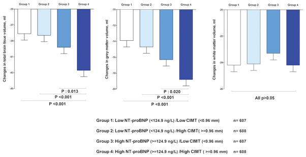
Approach and results: In the longitudinal population-based AGES-Reykjavik study (Age, Gene/Environment Susceptibility-Reykjavik), we included 2430 subjects (mean age, 74.6 years; 41.4% men) with baseline data on NT-proBNP and CITM (assessed by ultrasound imaging). Participants underwent a high-resolution brain magnetic resonance imaging at baseline and 5 years later to assess total brain (TBV), gray matter, and white matter volumes. Each unit higher log-transformed NT-proBNP was associated with 3.6 mL (95% confidence interval [CI], -6.0 to -1.1) decline in TBV and 3.5 mL (95% CI, -5.7 to -1.3) decline in gray matter volume. Likewise, each millimeter higher CIMT was associated with 10.8 mL (95% CI, -17.3 to -4.2) decline in TBV and 8.6 mL (95% CI, -14.4 to -2.8) decline in gray matter volume. There was no association between NT-proBNP and CIMT and changes in white matter volume. Compared with participants with low NT-proBNP and CIMT, participants with both high NT-proBNP and CIMT had 3.8 mL (95% CI, -6.0 to -1.6) greater decline in their TBV and 4 mL (95% CI, -6.0 to -2.0) greater decline in GMW. These associations were independent of sociodemographic and cardiovascular factors.
Conclusions: Older subjects with both cardiac dysfunction and carotid atherosclerosis are at an increased risk for brain parenchymal loss. Accumulated pathologies in the heart-brain axis might accelerate brain atrophy.
Keywords: brain; brain natriuretic peptide; carotid stenosis; gray matter; white matter.
© 2016 American Heart Association, Inc.
Figures
Comment in
-
Brain Atrophy, N-Terminal Brain Natriuretic Peptide, and Carotid Disease: Interconnecting Relationships Between Cerebral Perfusion, Cardiovascular Disease, Inflammation, and Cognitive Decline.Arterioscler Thromb Vasc Biol. 2016 Nov;36(11):2141-2142. doi: 10.1161/ATVBAHA.116.308362. Arterioscler Thromb Vasc Biol. 2016. PMID: 27784701 No abstract available.
Similar articles
-
N-terminal pro-brain natriuretic peptide and abnormal brain aging: The AGES-Reykjavik Study.Neurology. 2015 Sep 1;85(9):813-20. doi: 10.1212/WNL.0000000000001885. Epub 2015 Jul 31. Neurology. 2015. PMID: 26231259 Free PMC article.
-
Cross-Sectional Associations Between Cardiac Biomarkers, Cognitive Performance, and Structural Brain Changes Are Modified by Age.Arterioscler Thromb Vasc Biol. 2018 Aug;38(8):1948-1958. doi: 10.1161/ATVBAHA.118.311082. Arterioscler Thromb Vasc Biol. 2018. PMID: 29954754
-
NT-proBNP levels, atherosclerosis and vascular function in asymptomatic type 2 diabetic patients with microalbuminuria: peripheral reactive hyperaemia index but not NT-proBNP is an independent predictor of coronary atherosclerosis.Cardiovasc Diabetol. 2011 Aug 3;10:71. doi: 10.1186/1475-2840-10-71. Cardiovasc Diabetol. 2011. PMID: 21812947 Free PMC article.
-
NT-pro-B-type natriuretic peptide in infants and children: reference values based on combined data from four studies.Pediatr Cardiol. 2009 Jan;30(1):3-8. doi: 10.1007/s00246-008-9258-4. Epub 2008 Jul 4. Pediatr Cardiol. 2009. PMID: 18600369 Review.
-
Research digest: cardiac biomarkers for risk prediction.Lancet Diabetes Endocrinol. 2016 Nov;4(11):890. doi: 10.1016/S2213-8587(16)30293-5. Epub 2016 Oct 12. Lancet Diabetes Endocrinol. 2016. PMID: 27743976 Review. No abstract available.
Cited by
-
Cognitive Dysfunction after Heart Disease: A Manifestation of the Heart-Brain Axis.Oxid Med Cell Longev. 2021 Aug 18;2021:4899688. doi: 10.1155/2021/4899688. eCollection 2021. Oxid Med Cell Longev. 2021. PMID: 34457113 Free PMC article. Review.
-
N-Terminal pro-Brain Natriuretic Peptide and Associations With Brain Magnetic Resonance Imaging (MRI) Features in Middle Age: The CARDIA Brain MRI Study.Front Neurol. 2018 May 7;9:307. doi: 10.3389/fneur.2018.00307. eCollection 2018. Front Neurol. 2018. PMID: 29867721 Free PMC article.
-
Mobility-related brain regions linking carotid intima-media thickness to specific gait performances in old age.BMC Geriatr. 2024 Apr 1;24(1):303. doi: 10.1186/s12877-024-04918-1. BMC Geriatr. 2024. PMID: 38561655 Free PMC article.
-
Neurovascular-glymphatic dysfunction and white matter lesions.Geroscience. 2021 Aug;43(4):1635-1642. doi: 10.1007/s11357-021-00361-x. Epub 2021 Apr 14. Geroscience. 2021. PMID: 33851307 Free PMC article.
-
Transcranial sonography depicts a larger substantia nigra echogenic area in renal transplant patients on calcineurin inhibitors than on rapamycin.BMC Nephrol. 2022 Mar 17;23(1):108. doi: 10.1186/s12882-022-02741-7. BMC Nephrol. 2022. PMID: 35300603 Free PMC article.
References
-
- Knopman DS, Mosley TH, Catellier DJ, Sharrett AR Atherosclerosis Risk in Communities S. Cardiovascular risk factors and cerebral atrophy in a middle-aged cohort. Neurology. 2005;65:876–81. - PubMed
-
- Panerai RB. Cerebral autoregulation: from models to clinical applications. Cardiovascular engineering. 2008;8:42–59. - PubMed
-
- Vogels RL, van der Flier WM, van Harten B, Gouw AA, Scheltens P, Schroeder-Tanka JM, Weinstein HC. Brain magnetic resonance imaging abnormalities in patients with heart failure. European journal of heart failure. 2007;9:1003–9. - PubMed
Publication types
MeSH terms
Substances
Grants and funding
LinkOut - more resources
Full Text Sources
Other Literature Sources
Medical
Research Materials
