

doi: 10.14744/AnatolJCardiol.2016.7204.
Non-coronary abnormalities of the left heart: CT angiography findings
Affiliations
- PMID: 27609435
- PMCID: PMC5331359
- DOI: 10.14744/AnatolJCardiol.2016.7204
Item in Clipboard
Non-coronary abnormalities of the left heart: CT angiography findings
Anatol J Cardiol.
2016 Sep.
Abstract
Cardiac computed tomography (CT) is most commonly performed for the evaluation of the coronary arteries; however, non-coronary cardiac pathologies are frequently detected on these scans. In cases where magnetic resonance imaging cannot be used, cardiac CT can serve as the first-line imaging modality to evaluate many non-coronary cardiac pathologies. In this article, we discuss congenital non-coronary abnormalities of the left heart and their cardiac CT imaging features.
Conflict of interest statement
Figures

If a septal defect cannot be directly visualized due to its small size, a contrast jet (arrow) from the opacified left into the unenhanced right atrium will establish the diagnosis like in this patient

Primum atrial septal defect. Axial coronary CT angiography shows that the interatrial septum does not extend to the junction of the mitral and tricuspid valves, resulting in a defect (arrow) in the basal part of the septum. Note the thin septum in the fossa ovalis (arrow head) which should not be misinterpreted as a secundum ASD

A 68-year-old woman with secundum atrial septal defect. Axial coronary CT angiography image shows a large, incidentally detected defect (arrow) at the midportion of the interatrial septum
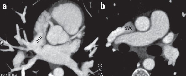
(a) Axial MIP coronary CT angiography image shows a connection (arrow) between two atria at the level of the opening of the superior vena cava (SVC) to the right atrium. (b) A right pulmonary vein drains into the SVC (known as partial anomalous pulmonary venous return) in the same patient

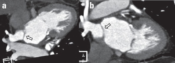
Coronary sinus type ASD. Normally, the coronary sinus (CS) is located along the posterior wall of the left atrium and drains into the right atrium (RA). In this case, MPR coronary CT angiography image shows a defect (arrow) between the roof of CS and left atrium (LA)

A variant of coronary sinus type ASD. (a) MPR coronary CT angiography image shows a defect (arrow) between the roof of coronary sinus and left atrium. (b) Axial coronary CT angiography image shows atresia of coronary sinus orifice (arrow head) in the same patient

(a, b) The patient with superior sinus venosus type ASD (arrow head) also has a secundum ASD (arrow). Cases with more than one type of ASD are called complex ASD

Patent foramen ovale. (a) Axial and (b) sagittal coronary CT angiography images show the lack of fusion between septum primum (black arrow) and secundum (white arrow) with a contrast-filled channel between them. The direction of the channel is towards the inferior vena cava (IVC). A contrast jet (arrow head) through the gap in the interatrial septum from the left to the right atrium is present

Coronary CT angiography image shows fusion of the septum primum and secundum in the inferior part (arrow head), and lack of fusion in the proximal part (arrow). In this case, a contrast-filled tunnel in the interatrial septum exists; however, a contrast jet to the right atrium does not occur. This entity is referred as probe patent foramen ovale

Interatrial septal aneurysm. (a) Coronary CT angiography image shows protrusion (more than 10 mm) of the interatrial septum into the right atrium. (b) In the same patient, a small defect (arrow) in the interatrial septum (secundum atrial septal defect) is also observed

(a) Coronary CT angiography image shows a diverticulum (black arrow) with a wide neck and smooth contours projecting outward from the inferior part of the left atrium wall. (b) Multiple left atrial diverticula (white arrows) are seen in a different patient

Coronary CT angiography image shows accessory left atrial appendage (arrow head) in addition to the normal left atrial appendage (arrow). Note the narrow neck and internal trabeculation of the accessory appendage

Coronary CT angiography image shows the co-existence of an accessory left atrial appendage (arrow) and left atrial diverticulum (arrow head) in the same patient. Left atrial diverticula have a wider neck and smoother contours compared to accessory left atrial appendages
Coronary CT angiography images show a thin membrane (arrows), which divides the left atrium into two compartments in a patient with cor triatriatum

Type 2 ventricular septal defect (VSD). Coronary CT angiography image shows a defect (arrow) in the membranous septum. This condition is referred as partially closed membranous VSD

Axial and sagittal coronary CT angiography images show protuberance of the interventricular septum into the right ventricle (arrows) in the location of the membranous septum. This condition is termed a ventricular septal aneurysm and is related to the spontaneous closure of a perimembranous ventricular septal defect

Type 3 (inlet type) ventricular septal defect (VSD). Coronary CT angiography image shows a VSD (arrow) at the inlet septum beneath the septal leaflet of the tricuspid valve (TV) (MV-mitral valve)

Type 4 ventricular septal defect. Coronary CT angiography image shows a defect (arrow) in the apical segment of the muscular interventricular septum

Coronary CT angiography image shows a ventricular cleft (arrow) in the interventricular septum of the left ventricle

Coronary CT angiography image shows multiple ventricular clefts (arrow) in the interventricular septum, which are connected to the left ventricle

Coronary CT angiography image shows a diverticulum (arrow) in the apical segment of the left ventricle in a patient with hypertrophic cardiomyopathy. A very thin left ventricular myocardium confining the diverticular sac is observable

Coronary CT angiography image shows that the ratio of non-compacted myocardium (NCM) to compacted myocardium (CM) is approximately 2.5 in the left ventricle in a patient with left ventricular non-compaction
Similar articles
-
German cardiac CT registry: indications, procedural data and clinical consequences in 7061 patients undergoing cardiac computed tomography.Int J Cardiovasc Imaging. 2018 May;34(5):807-819. doi: 10.1007/s10554-017-1282-0. Epub 2017 Dec 1. Int J Cardiovasc Imaging. 2018. PMID: 29197025
-
Fusion of CT coronary angiography and whole-heart dynamic 3D cardiac MR perfusion: building a framework for comprehensive cardiac imaging.Int J Cardiovasc Imaging. 2018 Apr;34(4):649-660. doi: 10.1007/s10554-017-1260-6. Epub 2017 Oct 28. Int J Cardiovasc Imaging. 2018. PMID: 29080955
-
Diagnostic accuracy of sub-mSv prospective ECG-triggering cardiac CT in young infant with complex congenital heart disease.Int J Cardiovasc Imaging. 2016 Jun;32(6):991-8. doi: 10.1007/s10554-016-0854-8. Epub 2016 Feb 20. Int J Cardiovasc Imaging. 2016. PMID: 26897005
-
Understanding the heart: CT and MRI for coronary heart disease.J Thorac Imaging. 2007 Feb;22(1):107-13. doi: 10.1097/RTI.0b013e3180317457. J Thorac Imaging. 2007. PMID: 17325582 Review.
-
Role of imaging studies in Kawasaki disease.Int J Rheum Dis. 2018 Jan;21(1):56-63. doi: 10.1111/1756-185X.13210. Epub 2017 Nov 8. Int J Rheum Dis. 2018. PMID: 29115035 Review.
Cited by
-
Inter-observer agreement of the Coronary Artery Disease Reporting and Data System (CAD-RADSTM) in patients with stable chest pain.Pol J Radiol. 2018 Apr 16;83:e151-e159. doi: 10.5114/pjr.2018.75641. eCollection 2018. Pol J Radiol. 2018. PMID: 30038693 Free PMC article.
-
Thickness and Volume of Epicardial Adipose Tissue in Relation to Stiffness and Elasticity of Aorta Assessed by Computed Tomography Angiography.Biomedicines. 2023 Jun 1;11(6):1617. doi: 10.3390/biomedicines11061617. Biomedicines. 2023. PMID: 37371711 Free PMC article.
-
Cross-sectional imaging findings of cardiac outpouchings.Diagn Interv Radiol. 2023 Jan 31;29(1):68-79. doi: 10.4274/dir.2022.221419. Epub 2023 Jan 2. Diagn Interv Radiol. 2023. PMID: 36960184 Free PMC article. Review.
-
Gamma-glutamyl transferase to albumin ratio can predict severity of coronary artery disease detected by coronary computed tomography angiography.Anatol J Cardiol. 2021 Sep;25(9):653-660. doi: 10.5152/AnatolJCardiol.2021.36330. Anatol J Cardiol. 2021. PMID: 34498597 Free PMC article.
References
-
- Chu LC, Johnson PT, Fishman EK. Cardiac CT angiography beyond the coronary arteries:what radiologists need to know and why they need to know it. AJR Am J Roengenol. 2014;203:583–95. - PubMed
-
- Brickner ME, Hillis D, Lange RA. Congenital heart disease in adults. First of two parts. N Eng J Med. 2000;342:256–63. - PubMed
-
- Navallas M, Orenes P, Sanchez Nistal MA, Jimenez Lopez Guarch C. Congenital heart disease in adults:The contribution of multidetector CT. Radiologia. 2010;52:288–300. - PubMed
-
- Hagen PT, Scholz DG, Edwards WD. Incidence and size of patent foramen ovale during the first 10 decades of life:An autopsy study of 965 normal hearts. Mayo Clin Proc. 1984;59:17–20. - PubMed
MeSH terms
LinkOut - more resources
Full Text Sources
Other Literature Sources
