

Structural Basis for High Specificity of Amadori Compound and Mannopine Opine Binding in Bacterial Pathogens
- PMID: 27609514
- PMCID: PMC5077200
- DOI: 10.1074/jbc.M116.745562
Structural Basis for High Specificity of Amadori Compound and Mannopine Opine Binding in Bacterial Pathogens
Abstract
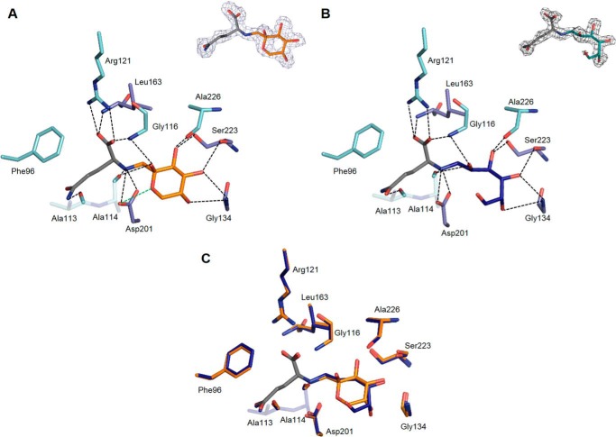
Agrobacterium tumefaciens pathogens genetically modify their host plants to drive the synthesis of opines in plant tumors. Opines are either sugar phosphodiesters or the products of condensed amino acids with ketoacids or sugars. They are Agrobacterium nutrients and imported into the bacterial cell via periplasmic-binding proteins (PBPs) and ABC-transporters. Mannopine, an opine from the mannityl-opine family, is synthesized from an intermediate named deoxy-fructosyl-glutamine (DFG), which is also an opine and abundant Amadori compound (a name used for any derivative of aminodeoxysugars) present in decaying plant materials. The PBP MotA is responsible for mannopine import in mannopine-assimilating agrobacteria. In the nopaline-opine type agrobacteria strain, SocA protein was proposed as a putative mannopine binding PBP, and AttC protein was annotated as a mannopine binding-like PBP. Structural data on mannityl-opine-PBP complexes is currently lacking. By combining affinity data with analysis of seven x-ray structures at high resolution, we investigated the molecular basis of MotA, SocA, and AttC interactions with mannopine and its DFG precursor. Our work demonstrates that AttC is not a mannopine-binding protein and reveals a specific binding pocket for DFG in SocA with an affinity in nanomolar range. Hence, mannopine would not be imported into nopaline-type agrobacteria strains. In contrast, MotA binds both mannopine and DFG. We thus defined one mannopine and two DFG binding signatures. Unlike mannopine-PBPs, selective DFG-PBPs are present in a wide diversity of bacteria, including Actinobacteria, α-,β-, and γ-proteobacteria, revealing a common role of this Amadori compound in pathogenic, symbiotic, and opportunistic bacteria.
Keywords: ABC transporter; DFG; crystal structure; host-pathogen interaction; isothermal titration calorimetry (ITC); mannopine; opine; periplasmic binding protein; plant pathogen; x-ray crystallography.
© 2016 by The American Society for Biochemistry and Molecular Biology, Inc.
Figures


Similar articles
-
Import pathways of the mannityl-opines into the bacterial pathogen Agrobacterium tumefaciens: structural, affinity and in vivo approaches.Biochem J. 2020 Feb 14;477(3):615-628. doi: 10.1042/BCJ20190886. Biochem J. 2020. PMID: 31922182
-
Structural basis for two efficient modes of agropinic acid opine import into the bacterial pathogen Agrobacterium tumefaciens.Biochem J. 2019 Jan 15;476(1):165-178. doi: 10.1042/BCJ20180861. Biochem J. 2019. PMID: 30552142
-
Convergent evolution of Amadori opine catabolic systems in plasmids of Agrobacterium tumefaciens.J Bacteriol. 2003 Jan;185(2):513-24. doi: 10.1128/JB.185.2.513-524.2003. J Bacteriol. 2003. PMID: 12511498 Free PMC article.
-
[Opine biosynthesis and catabolism genes of Agrobacterium tumefaciens and Agrobacterium rhizogenes].Genetika. 2015 Feb;51(2):137-46. Genetika. 2015. PMID: 25966579 Review. Russian.
-
Opine biosynthesis in naturally transgenic plants: Genes and products.Phytochemistry. 2021 Sep;189:112813. doi: 10.1016/j.phytochem.2021.112813. Epub 2021 Jun 27. Phytochemistry. 2021. PMID: 34192603 Review.
Cited by
-
Tobacco Root Endophytic Arthrobacter Harbors Genomic Features Enabling the Catabolism of Host-Specific Plant Specialized Metabolites.mBio. 2021 Jun 29;12(3):e0084621. doi: 10.1128/mBio.00846-21. Epub 2021 May 28. mBio. 2021. PMID: 34044592 Free PMC article.
-
The rhizobial type III effector ErnA confers the ability to form nodules in legumes.Proc Natl Acad Sci U S A. 2019 Oct 22;116(43):21758-21768. doi: 10.1073/pnas.1904456116. Epub 2019 Oct 7. Proc Natl Acad Sci U S A. 2019. PMID: 31591240 Free PMC article.
-
Multicentered hydrogen bonding in 1-[(1-de-oxy-β-d-fructo-pyranos-1-yl)aza-nium-yl]cyclo-pentane-carboxyl-ate ('d-fructose-cyclo-leucine').Acta Crystallogr E Crystallogr Commun. 2019 Jul 2;75(Pt 8):1096-1101. doi: 10.1107/S2056989019009253. eCollection 2019 Aug 1. Acta Crystallogr E Crystallogr Commun. 2019. PMID: 31417772 Free PMC article.
-
Ecological Conditions and Molecular Determinants Involved in Agrobacterium Lifestyle in Tumors.Front Plant Sci. 2019 Jul 30;10:978. doi: 10.3389/fpls.2019.00978. eCollection 2019. Front Plant Sci. 2019. PMID: 31417593 Free PMC article. Review.
-
The crystal structure of the Yersinia pestis iron chaperone YiuA reveals a basic triad binding motif for the chelated metal.Acta Crystallogr D Struct Biol. 2017 Nov 1;73(Pt 11):921-939. doi: 10.1107/S2059798317015236. Epub 2017 Oct 26. Acta Crystallogr D Struct Biol. 2017. PMID: 29095164 Free PMC article.
References
-
- Schell J., Van Montagu M., De Beuckeleer M., De Block M., Depicker A., De Wilde M., Engler G., Genetello C., Hernalsteens J. P., Holsters M., Seurinck J., Silva B., Van Vliet F., and Villarroel R. (1979) Interactions and DNA transfer between Agrobacterium tumefaciens, the Ti-plasmid, and the plant host. Proc. R. Soc. Lond. Ser. B Biol. Sci. 204, 251–266 - PubMed
-
- Tempé J., and Petit A. (1983) La piste des opines. In Molecular Genetics of the Bacteria-Plant Interaction (Pühler A. ed), pp. 14–32, Berlin-Heidelberg, Springer-Verlag
-
- Dessaux Y., Petit A., and Tempe J. (1993) Chemistry and biochemistry of opines, chemical mediators of parasitism. Phytochemistry 34, 31–38
MeSH terms
Substances
Associated data
- Actions
- Actions
- Actions
- Actions
- Actions
- Actions
- Actions
- Actions
- Actions
- Actions
- Actions
- Actions
LinkOut - more resources
Full Text Sources
Other Literature Sources
Research Materials
Miscellaneous
