

Prenatal Evaluation, Imaging Features, and Neurodevelopmental Outcome of Prenatally Diagnosed Periventricular Pseudocysts
- PMID: 27609618
- PMCID: PMC7963870
- DOI: 10.3174/ajnr.A4916
Prenatal Evaluation, Imaging Features, and Neurodevelopmental Outcome of Prenatally Diagnosed Periventricular Pseudocysts
Abstract
Background and purpose: Periventricular pseudocysts are cystic cavities that lack the ependymal cell lining found in true cysts. The aim of this study was to characterize periventricular pseudocysts and related findings and their neurodevelopmental outcome.
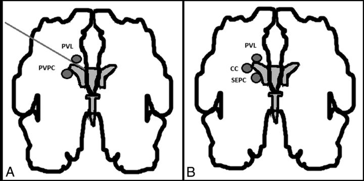
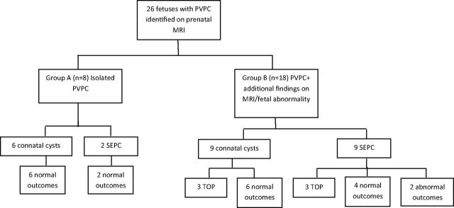
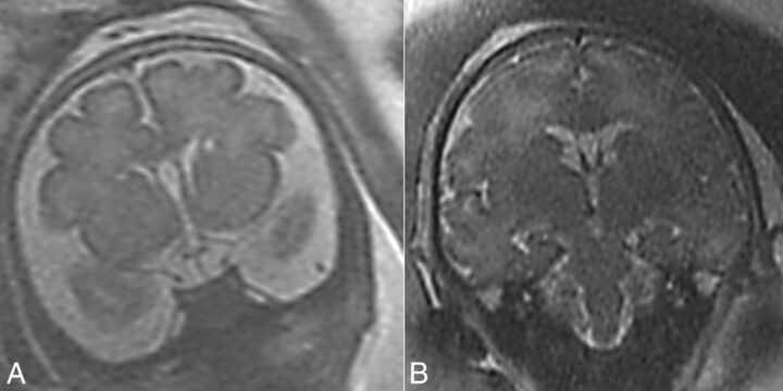
Materials and methods: This was a retrospective study of periventricular pseudocysts detected prenatally on fetal MR imaging in 26 fetuses. The fetuses were divided into group A (n = 8), which included cases with isolated periventricular pseudocysts, and group B (n = 18), which included cases of periventricular pseudocysts with additional findings. Cases were further subdivided into connatal cysts and subependymal pseudocysts. Data collected included prenatal history, MR imaging features, sonographic follow-up, and neurodevelopmental outcome.
Results: All cases in group A (n = 8) had a normal outcome. In group B (n = 18), 6 pregnancies were terminated and 2 had an abnormal outcome. Both cases with an abnormal outcome involved patients with subependymal pseudocysts. No significant association was found between the morphologic features on MR imaging and the neurodevelopmental outcome.
Conclusions: Neurodevelopmental outcome in cases of isolated periventricular pseudocysts detected prenatally appears to be normal. A detailed evaluation should be performed to rule out additional brain findings, chromosomal aberration, and fetal malformation. This evaluation should include the following: maternal TORCH status, detailed fetal sonographic anatomic evaluation, fetal echocardiogram, fetal brain MR imaging, amniocentesis and karyotyping/comparative genomic hybridization, and genetic counseling. Additional findings on MR imaging, including mild-to-moderate dilated ventricles, asymmetric ventricles, or T2 hyperintense signal in the white matter without other findings or major fetal abnormality, appear to be benign. Connatal cysts appear to be benign.
© 2016 by American Journal of Neuroradiology.
Figures
Similar articles
-
Congenital periventricular pseudocysts: prenatal sonographic appearance and clinical implications.Ultrasound Obstet Gynecol. 2002 Nov;20(5):447-51. doi: 10.1046/j.1469-0705.2002.00840.x. Ultrasound Obstet Gynecol. 2002. PMID: 12423480
-
Connatal periventricular pseudocysts in the neonate.Pediatr Radiol. 1992;22(1):55-8. doi: 10.1007/BF02011609. Pediatr Radiol. 1992. PMID: 1594311
-
Prenatal diagnosis of arachnoid cysts: MRI features and neurodevelopmental outcome.Eur J Radiol. 2019 Apr;113:232-237. doi: 10.1016/j.ejrad.2019.02.027. Epub 2019 Feb 22. Eur J Radiol. 2019. PMID: 30927952
-
Subependymal pseudocysts in the fetal brain: prenatal diagnosis of two cases and review of the literature.Ultrasound Obstet Gynecol. 2002 Nov;20(5):502-5. doi: 10.1046/j.1469-0705.2002.00848.x. Ultrasound Obstet Gynecol. 2002. PMID: 12423490 Review.
-
Mild fetal ventriculomegaly: diagnosis, evaluation, and management.Am J Obstet Gynecol. 2018 Jul;219(1):B2-B9. doi: 10.1016/j.ajog.2018.04.039. Epub 2018 Apr 26. Am J Obstet Gynecol. 2018. PMID: 29705191 Review.
Cited by
-
MRI Findings at Term-Corrected Age and Neurodevelopmental Outcomes in a Large Cohort of Very Preterm Infants.AJNR Am J Neuroradiol. 2020 Aug;41(8):1509-1516. doi: 10.3174/ajnr.A6666. AJNR Am J Neuroradiol. 2020. PMID: 32796100 Free PMC article.
-
Association between White Matter T2 Hyper-Intense Signals in Fetal Brain Magnetic Resonance Imaging and Neurodevelopment of Fetuses with Cytomegalovirus Infection.Diagnostics (Basel). 2024 Apr 11;14(8):797. doi: 10.3390/diagnostics14080797. Diagnostics (Basel). 2024. PMID: 38667443 Free PMC article.
-
Evaluation of MRI Features and Neurodevelopmental Outcomes for Prenatally Diagnosed Periventricular Pseudocysts.Front Pediatr. 2021 Oct 22;9:681999. doi: 10.3389/fped.2021.681999. eCollection 2021. Front Pediatr. 2021. PMID: 34746043 Free PMC article.
-
The Lateral Ventricles: A Detailed Review of Anatomy, Development, and Anatomic Variations.AJNR Am J Neuroradiol. 2020 Apr;41(4):566-572. doi: 10.3174/ajnr.A6456. Epub 2020 Feb 20. AJNR Am J Neuroradiol. 2020. PMID: 32079598 Free PMC article. Review.
-
A case of Aicardi-Goutières syndrome caused by TREX1 gene mutation.BMC Pregnancy Childbirth. 2023 Feb 22;23(1):124. doi: 10.1186/s12884-023-05436-5. BMC Pregnancy Childbirth. 2023. PMID: 36814213 Free PMC article.
References
MeSH terms
LinkOut - more resources
Full Text Sources
Other Literature Sources
Medical