. 2016 Aug 25:7:1255.
doi: 10.3389/fpls.2016.01255. eCollection 2016.

Photosynthate Regulation of the Root System Architecture Mediated by the Heterotrimeric G Protein Complex in Arabidopsis

Affiliations

Photosynthate Regulation of the Root System Architecture Mediated by the Heterotrimeric G Protein Complex in Arabidopsis

Yashwanti Mudgil et al. Front Plant Sci. .

Abstract

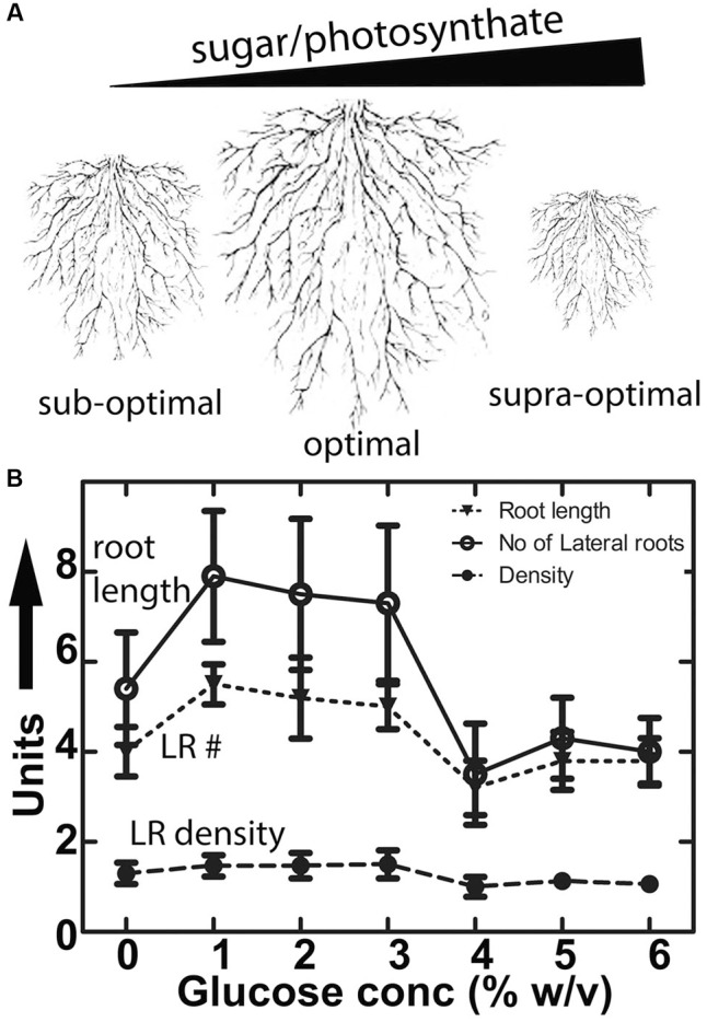
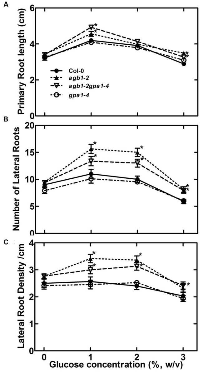
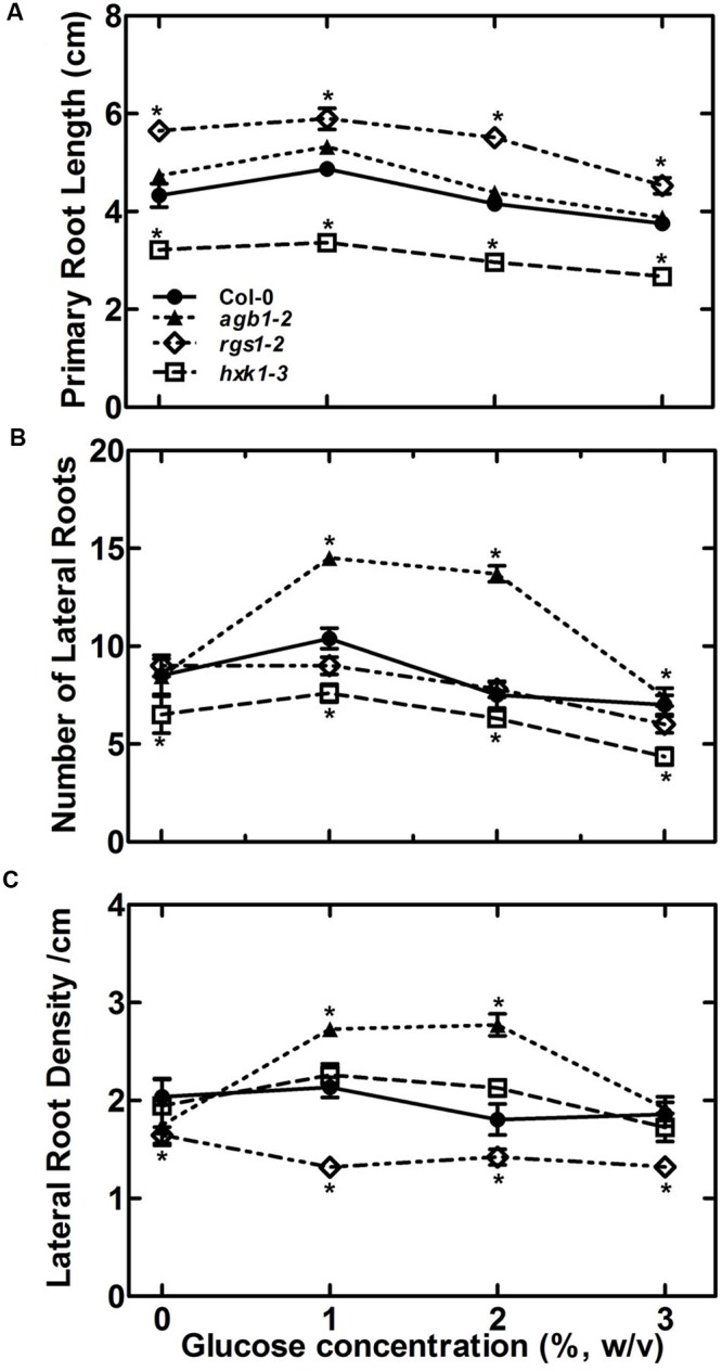
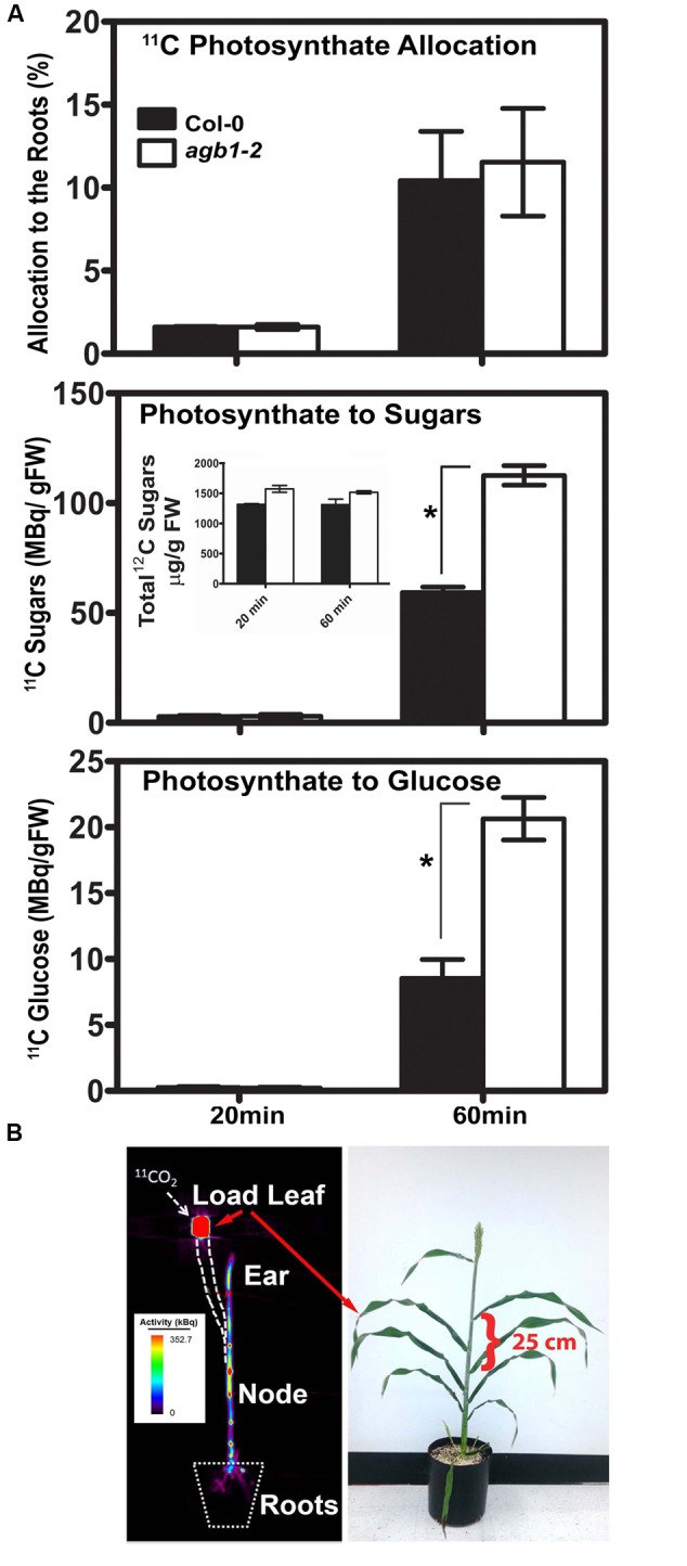
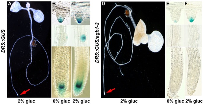
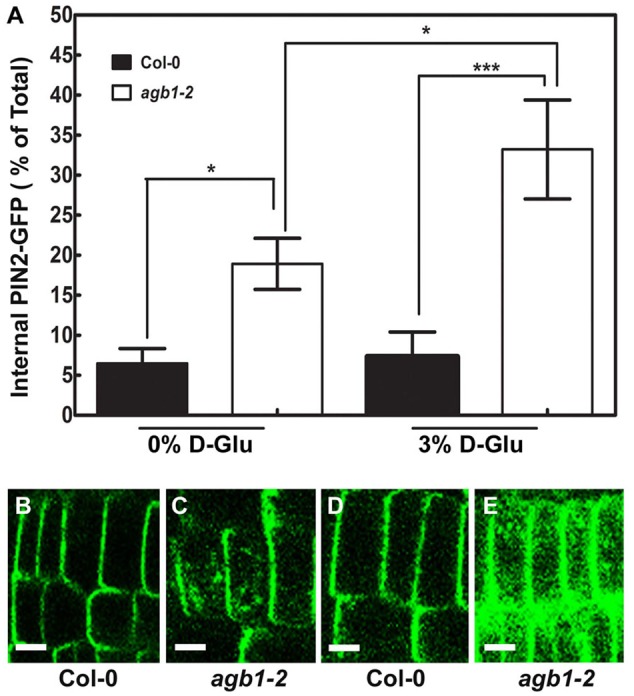
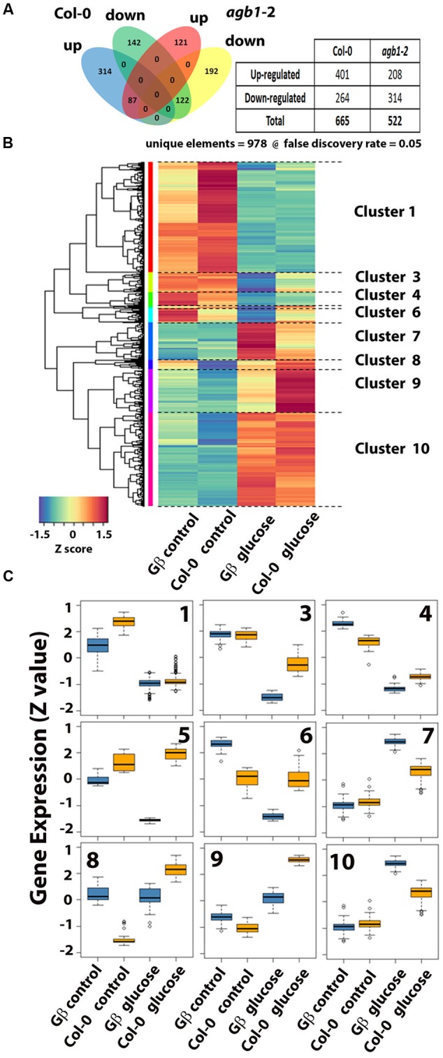
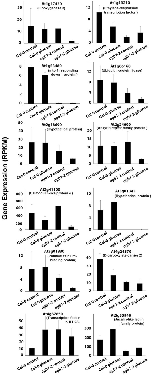
Assimilate partitioning to the root system is a desirable developmental trait to control but little is known of the signaling pathway underlying partitioning. A null mutation in the gene encoding the Gβ subunit of the heterotrimeric G protein complex, a nexus for a variety of signaling pathways, confers altered sugar partitioning in roots. While fixed carbon rapidly reached the roots of wild type and agb1-2 mutant seedlings, agb1 roots had more of this fixed carbon in the form of glucose, fructose, and sucrose which manifested as a higher lateral root density. Upon glucose treatment, the agb1-2 mutant had abnormal gene expression in the root tip validated by transcriptome analysis. In addition, PIN2 membrane localization was altered in the agb1-2 mutant. The heterotrimeric G protein complex integrates photosynthesis-derived sugar signaling incorporating both membrane-and transcriptional-based mechanisms. The time constants for these signaling mechanisms are in the same range as photosynthate delivery to the root, raising the possibility that root cells are able to use changes in carbon fixation in real time to adjust growth behavior.

Keywords: AGB1; PIN2-GFP; gene expression; glucose; lateral root density; photosynthetic partitioning; positron electron tomography imaging.


Figures

FIGURE 1
FIGURE 1
Conserved architecture of plant root growth in nature and the lab. (A) Diagrammatic representation of roots of different size showing constant lateral root density and lateral root primordial position grown under sub-optimal, optimal and supra-optimal nutrient conditions. (B) Primary root length (mm), number of lateral roots (per root) and lateral root density (roots per mm) in Col-0 in the presence of different concentrations of glucose. Values represent the mean of 3 independent experiments (n = 10 each); bars represent the standard error.
FIGURE 2
FIGURE 2
Role of G protein subunits in sensing sugar in RSA maintenance. (A) Primary root length of 11-day-old seedlings of Gα, Gβ and Gαβ double subunit mutants (indicated genotypes) were grown on ½ X MS, and 0.75% agar, 22°C, 8:16 light: dark cycle for 4 days followed by 7 days of vertical growth on different concentrations of glucose. (B) Lateral root number (C) Lateral root density. All experiments were repeated 3 times with 10–15 seedlings of each genotype per trial. Error bars represent standard error Student’s t-test results are based on difference between the wild type and indicated genotype shown as asterisks:P < 0.05.
FIGURE 3
FIGURE 3
The sugar sensing mechanism in RSA maintenance may involve both HEXOKINASE 1(HXK1) and REGULATOR of G SIGNALING 1 (RGS1). Effect of glucose on RSA in terms of primary root length (A), number of lateral roots (B) and lateral root density (C) was compared for wild type, agb1-2, hxk1-3 and rgs1-2 mutant. All experiments were repeated three times with 10–15 seedlings used for each genotype in each trial. Error bars represent standard error Student’s t-test results are based on difference between the wild type and indicated genotype shown as asterisks: P < 0.05.
FIGURE 4
FIGURE 4
Positron electron tomography imaging of allocation and partitioning of photoassimilate. (A) Top panel. Total allocation of [11C] CO2 in the roots of Col (solid bars) and agb1-2 (open bars) at the indicated times. Middle panel. Partitioning of newly fixed 11CO2 to soluble sugars (glucose, fructose and sucrose) in the roots of Col (solid bars) and agb1-2 (open bars) at the indicated times. (Inset) Total non-radioactive [12C] sugars (glucose, fructose and sucrose) in the roots of Col (solid bars) and agb1-2 (open bars) at the indicated times. Non-radioactive [12C] sugars were extracted and analyzed by thin layer chromatography as described previously (Babst et al., 2013). Numbers represent the average of 3 independent experiments (n = 10 each) and error bars represent SE. Lower panel. Partitioning of newly fixed 11CO2 to glucose in the roots. Percentage values were calculated as radioactivity in the roots relative to the total seedling activity. Radioactivity (MBq/g FW) represent the mean of 3 independent experiments (n = 10 each) and error bars represent the standard error. (B) Fixed carbon is rapidly distributed to tissue sinks. (Left panel) The image shows the distribution of 11C-labeled photoassimilate in different parts of in an intact sorghum plant shown in the right panel imaged 2 h after 11CO2 administration to the load leaf at the position indicated. Heat scale represents activity/pixel. Load leaf = site of 11CO2 administration. Velocity was 1.25 cm min-1. A distance of 25 cm is indicated by the bracket. Student’s t-test results are based on difference between the means. Asterisks indicate P < 0.001.
FIGURE 5
FIGURE 5
Glucose-induced auxin maxima in agb1-2 mutant roots. DR5::GUS is a synthetic, auxin-inducible gene promoter reporter used to detect auxin maxima or auxin signaling. (A–C) Wild type and agb1-2 (D–F) seedlings were treated with 2% glucose (gluc) for 4 h (A,C,D,F) and compared to the untreated controls (B,E). Arrows point to the root tip. Compared to wild type, glucose did not increase DR5-driven expression of GUS in the agb1-2 root tips (cf. B,C; lower panels to E,F; lower panels) and lateral root primordial (cf. B,C; middle panels to E,F; middle panels), although DR5::GUS expression occurred in emergent lateral roots (cf. B,C; upper panels to E,F; upper panels).
FIGURE 6
FIGURE 6
Altered subcellular localization of PIN2-GFP in the agb1-2 mutant. (A) Quantitation of PIN2-GFP subcellular localization in Col-0 and agb1-2 root tip cells grown with or without supplemental 3% glucose. Fluorescence intensities in multiple seedlings were measured using ImageJ software and compared using Student’s t-test, shown as asterisks:P < 0.05; ∗∗∗P < 0.0005. PIN2-GFP localization in Col-0 root tip cells (B,D) grown without (-) or with (+) supplemental 3% glucose, respectively. (C,E) Amount of PIN2-GFP internalized in agb1-2 root tip cells grown without (-) or with (+) supplemental 3% glucose, respectively. Scale bars represent 5 μm. In order to compare directly these genotypes, PIN2-GFP/agb1-2 lines were obtained by crossing agb1-2 plants into the PIN2-GFP line shown in panels B and D. This experiment was repeated three times and reproducible PIN2-GFP localization pattern was observed upon glucose treatment.
FIGURE 7
FIGURE 7
Glucose-induced gene expression in the 1-mm root tips of wild type and agb1-2 mutant. Five-day old, etiolated seedlings were treated with glucose for 4 h and the apical 1-mm of roots was harvested for RNA profiling as described in Materials and Methods. (A) Venn diagram quantitating genes that were differentially expressed in the two genotypes. The table inset summarizes the number of genes scored as up or down regulated in each genotype. (B) Cluster analysis of the genes that have altered expression displayed as a heat map. Ten distinct clusters were formed. Clusters 2 and 5 had too few genes to label. (C) The expression profile of each cluster is shown as a box plot. The top of the rectangle indicates the third quartile, the horizontal line indicates the median, and the bottom of the rectangle indicates the first quartile. The vertical line from the top indicates the maximum value, and the other vertical line extending from the bottom indicates the minimum value.
FIGURE 8
FIGURE 8
Glucose regulation of genes that differ between wild type and agb1-2 root tips. Genes that are differentially expressed (FDR ≤ 0.05) between the two genotypes in at least one of the conditions (control and glucose treatment) are shown. Normalized gene expression is shown as reads per kb per million reads (RPKM).

Similar articles

Cited by

References

    1. Albrechtova J., Kubinova Z., Soukup A., Janacek J. (2014). Image analysis: basic procedures for description of plant structures. Methods Mol. Biol. 1080 67–76. 10.1007/978-1-62703-643-6_5 - DOI - PubMed
    1. Anders S., Pyl P., Huber W. (2014). HTSeq-A Python framework to work with high-throughput sequencing data. Bioinformatics 31 166–169. 10.1093/bioinformatics/btu638 - DOI - PMC - PubMed
    1. Babst B. A., Karve A. A., Judt T. (2013). Radio-metabolite analysis of carbon-11 biochemical partitioning to non-structural carbohydrates for integrated metabolism and transport studies. Plant Cell Physiol. 54 1016–1025. 10.1093/pcp/pct045 - DOI - PubMed
    1. Barbier F. F., Lunn J. E., Beveridge C. A. (2015). Ready, steady, go! A sugar hit starts the race to shoot branching. Curr. Opin. Plant Biol. 25 39–45. 10.1016/j.pbi.2015.04.004 - DOI - PubMed
    1. Benková E., Michniewicz M., Sauer M., Teichmann T., Seifertová D., Jürgens G., et al. (2003). Local, efflux-dependent auxin gradients as a common module for plant organ formation. Cell 115 591–602. 10.1016/S0092-8674(03)00924-3 - DOI - PubMed

LinkOut - more resources