. 2016 Aug 1;5(1):235-48.
doi: 10.1089/biores.2016.0027. eCollection 2016.

Hepatic Differentiation of Human Induced Pluripotent Stem Cells in a Perfused Three-Dimensional Multicompartment Bioreactor

Affiliations

Hepatic Differentiation of Human Induced Pluripotent Stem Cells in a Perfused Three-Dimensional Multicompartment Bioreactor

Nora Freyer et al. Biores Open Access. .

Abstract

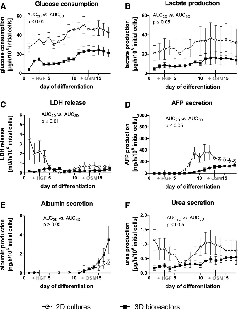
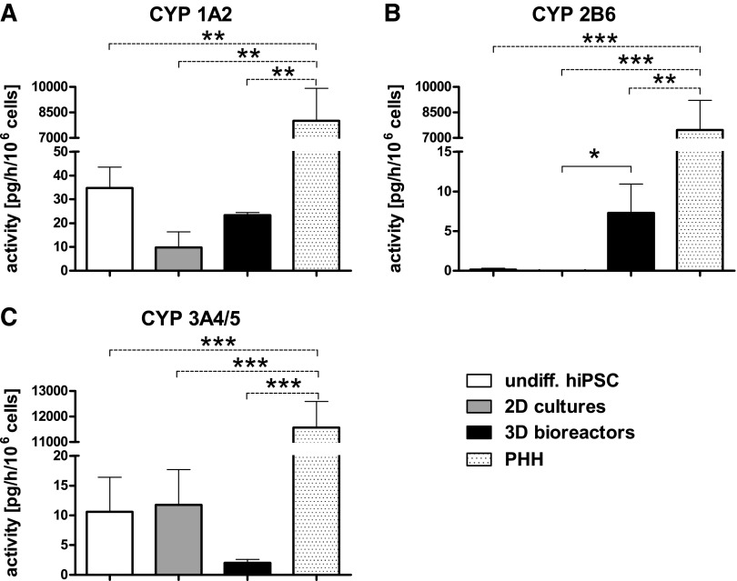
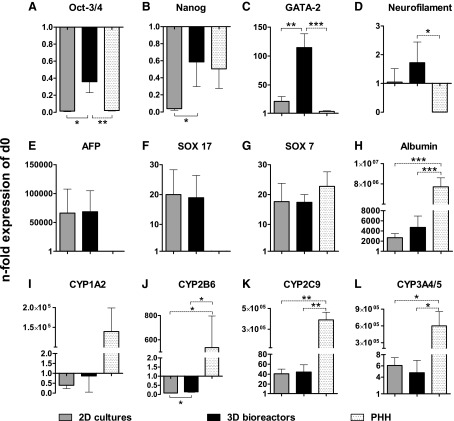
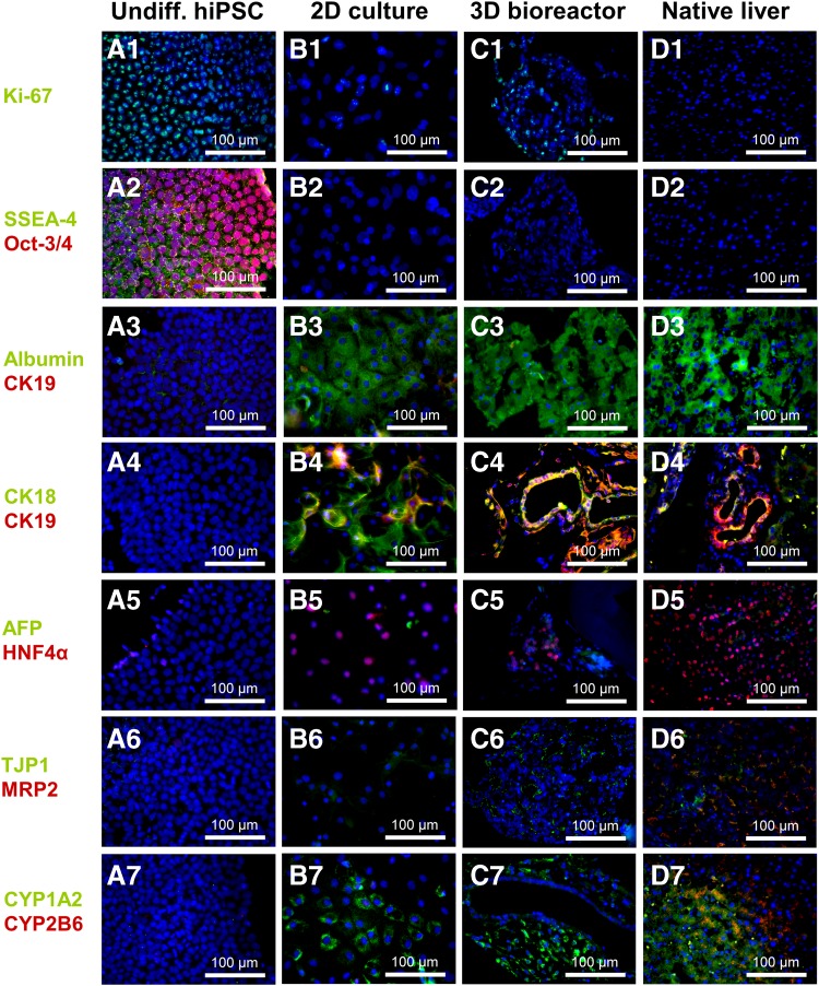
The hepatic differentiation of human induced pluripotent stem cells (hiPSC) holds great potential for application in regenerative medicine, pharmacological drug screening, and toxicity testing. However, full maturation of hiPSC into functional hepatocytes has not yet been achieved. In this study, we investigated the potential of a dynamic three-dimensional (3D) hollow fiber membrane bioreactor technology to improve the hepatic differentiation of hiPSC in comparison to static two-dimensional (2D) cultures. A total of 100 × 10(6) hiPSC were seeded into each 3D bioreactor (n = 3). Differentiation into definitive endoderm (DE) was induced by adding activin A, Wnt3a, and sodium butyrate to the culture medium. For further maturation, hepatocyte growth factor and oncostatin M were added. The same differentiation protocol was applied to hiPSC maintained in 2D cultures. Secretion of alpha-fetoprotein (AFP), a marker for DE, was significantly (p < 0.05) higher in 2D cultures, while secretion of albumin, a typical characteristic for mature hepatocytes, was higher after hepatic differentiation of hiPSC in 3D bioreactors. Functional analysis of multiple cytochrome P450 (CYP) isoenzymes showed activity of CYP1A2, CYP2B6, and CYP3A4 in both groups, although at a lower level compared to primary human hepatocytes (PHH). CYP2B6 activities were significantly (p < 0.05) higher in 3D bioreactors compared with 2D cultures, which is in line with results from gene expression. Immunofluorescence staining showed that the majority of cells was positive for albumin, cytokeratin 18 (CK18), and hepatocyte nuclear factor 4-alpha (HNF4A) at the end of the differentiation process. In addition, cytokeratin 19 (CK19) staining revealed the formation of bile duct-like structures in 3D bioreactors similar to native liver tissue. The results indicate a better maturation of hiPSC in the 3D bioreactor system compared to 2D cultures and emphasize the potential of dynamic 3D culture systems in stem cell differentiation approaches for improved formation of differentiated tissue structures.

Keywords: stem cells; tissue engineering.


Figures

<b>FIG. 1.</b>
FIG. 1.
Metabolic activity of hiPSC during hepatic differentiation in 2D cultures (dotted line) or in 3D bioreactor cultures (black line). (A) Glucose consumption, (B) lactate production, (C) release of LDH, (D) secretion of AFP, (E) albumin production, and (F) urea secretion. Values were normalized to 1 × 106 inoculated cells. AUC was calculated and differences were detected with the unpaired, two-tailed Student's t-test (3D bioreactors: n = 3, 2D cultures: n = 4, mean ± SEM). AFP, alpha-fetoprotein; AUC, area under curve; 2D, two dimensional; 3D, three dimensional; hiPSC, human induced pluripotent stem cells; LDH, lactate dehydrogenase; SEM, standard error of the mean.
<b>FIG. 2.</b>
FIG. 2.
Activities of different cytochrome P450 (CYP) isoenzymes in undifferentiated hiPSC (white), in hiPSC after hepatic differentiation in 2D cultures (gray) or 3D bioreactors (black), or in PHH (dotted). CYP activities were determined by measuring the conversion rates of selected substrates into isoenzyme-specific products. (A) Formation of acetaminophen from phenacetin by CYP1A2, (B) formation of 6-OH-bupropion from bupropion by CYP2B6, and (C) formation of 1-OH-midazolam from midazolam by CYP3A4/5. Differences in metabolic activity between undifferentiated hiPSC, 2D cultures and 3D bioreactors, were calculated using one-way ANOVA with Bonferroni's multiple comparison test (solid line). In addition, differences between PHH to all other groups were calculated using one-way ANOVA with Dunnett's multiple comparison test (dotted line). (3D bioreactors: n = 3, 2D cultures and undifferentiated hiPSC: n = 4, PHH: n = 5; mean ± SEM), *p ≤ 0.05, **p ≤ 0.01, ***p ≤ 0.001. ANOVA, analysis of variance; PHH, primary human hepatocytes.
<b>FIG. 3.</b>
FIG. 3.
Gene expression of pluripotency markers (A) Oct-3/4 and (B) Nanog, mesodermal marker (C) GATA-2, ectodermal marker (D) neurofilament, endodermal markers (E) AFP and (F) SOX 17, extra-embryonic marker (G) SOX 7 and hepatic markers (H) albumin, (I) CYP1A2, (J) CYP2B6, (K) CYP2C9 and (L) CYP3A4/5 in hiPSC after hepatic differentiation in 2D cultures or 3D bioreactors, and in PHH relative to undifferentiated hiPSC (d0). Samples for mRNA expression analysis were taken after hepatic differentiation of hiPSC in 2D cultures (black) or 3D bioreactors (gray). For mRNA expression analysis of PHH (dotted), freshly isolated cells were used. Fold changes relative to undifferentiated hiPSC were calculated with normalization to GAPDH expression by the ΔΔCt method. Differences in gene expression between 2D cultures and 3D bioreactors were calculated using the unpaired, two-tailed Student's t-test (solid line). In addition, differences between PHH and all other groups were calculated by means of one-way ANOVA with Dunnett's multiple comparison test (dotted line) (3D bioreactors: n = 3, 2D cultures and undifferentiated hiPSC: n = 4, PHH: n = 3; mean ± SEM), *p ≤ 0.05, **p ≤ 0.01, ***p ≤ 0.001.
<b>FIG. 4.</b>
FIG. 4.
Immunofluorescence analysis of hiPSC before and after hepatic differentiation in 2D cultures or 3D bioreactors compared with native human liver tissue. Samples from cultures or liver tissue were stained with (A1–D1) Ki-67, (A2–D2) SSEA-4 and Oct-3/4, (A3–D3) albumin and CK19, (A4–D4) CK18 and CK19, (A5–D5) AFP and HNF4α, (A6–D6) TJP1 and MRP2, and with (A7–D7) CYP1A2 and CYP2B6. Nuclei were counterstained with DAPI (blue) or with bisBenzimide H 33342 trihydrochloride in native human liver tissue (blue). CK19, cytokeratin 19.
<b>FIG. 5.</b>
FIG. 5.
Ultrastructural characteristics of hiPSC after hepatic differentiation in 3D bioreactors. (A) Cells with a high nucleus:cytoplasm ratio indicating immature cells (N). (B) Cells with distinct microvilli (Mv) and abundant cell–cell contacts (arrows) between neighboring cells. (C) Tight junction (TJ) and desmosome (De) between two neighboring cells. (D) Interdigitations (Id) between two cells with mitochondria (M), rough endoplasmic reticulum (rER), and Golgi apparatus (G).

References

    1. Kola I, Landis J. Can the pharmaceutical industry reduce attrition rates? Nat Rev Drug Discov. 2004;3:711–715 - PubMed
    1. Lewis DF, Ioannides C, Parke DV. Cytochromes P450 and species differences in xenobiotic metabolism and activation of carcinogen. Environ Health Perspect. 1998;106:633–641 - PMC - PubMed
    1. Giri S, Bader A. Improved preclinical safety assessment using micro-BAL devices: the potential impact on human discovery and drug attrition. Drug Discov Today. 2011;16:382–397 - PubMed
    1. Ingelman-Sundberg M. Pharmacogenetics of cytochrome P450 and its applications in drug therapy: the past, present and future. Trends Pharmacol Sci. 2004;25:193–200 - PubMed
    1. Takayama K, Morisaki Y, Kuno S, et al. . Prediction of interindividual differences in hepatic functions and drug sensitivity by using human iPS-derived hepatocytes. Proc Natl Acad Sci U S A. 2014;111:16772–16777 - PMC - PubMed

LinkOut - more resources