

History of the infantile hepatic hemangioma: From imaging to generating a differential diagnosis
- PMID: 27610342
- PMCID: PMC4978619
- DOI: 10.5409/wjcp.v5.i3.273
History of the infantile hepatic hemangioma: From imaging to generating a differential diagnosis
Abstract
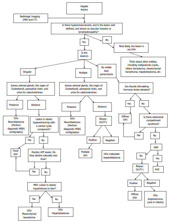
We aim to provide an up-to-date summary of infantile hepatic hemangioma (IHH) and its misnomers and to dialectically present the differential diagnosis of these rare entities of the liver. Eligible peer-reviewed articles on hepatic infantile hemangiomas, published between 2000 and 2015, were reviewed for this study. IHH is the most common hepatic vascular tumor in children. Once a liver mass is identified in an infant, the differential diagnosis ranges from vascular malformations to benign and malignant tumors including mesenchymal hamartoma, hepatoblastoma, metastatic neuroblastoma, so careful physical examination, imaging studies, and, if indicated, tumor markers and biopsy, are of pivotal importance to ascertain the correct diagnosis. Despite the benign nature of IHHs, some of these lesions may demand medical and/or surgical intervention, especially for multiple and diffuse IHH. Complications can include hepatomegaly, hypothyroidism and cardiac failure. Therefore, a close follow-up is required until complete involution of the lesions. We propose an algorithm to guide the physicians towards the proper management of hepatic lesions.
Keywords: Children; Hepatic hemangiomas; Infant; Vascular tumors.
Figures
References
-
- Drolet BA, Swanson EA, Frieden IJ. Infantile hemangiomas: an emerging health issue linked to an increased rate of low birth weight infants. J Pediatr. 2008;153:712–715, 715.e1. - PubMed
-
- Patiño-Seijas B, Lorenzo-Franco F, Rey-Sanjurjo JL, González-Cuesta M, López-Cedrún Cembranos JL. Vascular Lesions: GLUT-1 expression as a diagnostic tool to discriminate tumors from malformations. J Oral Maxillofac Surg. 2012;70:2333–2342. - PubMed
-
- Dickie B, Dasgupta R, Nair R, Alonso MH, Ryckman FC, Tiao GM, Adams DM, Azizkhan RG. Spectrum of hepatic hemangiomas: management and outcome. J Pediatr Surg. 2009;44:125–133. - PubMed
-
- Lu CC, Ko SF, Liang CD, Kuo HW, Tiao MM. Infantile hepatic hemangioendothelioma presenting as early heart failure: report of two cases. Chang Gung Med J. 2002;25:405–410. - PubMed
Publication types
LinkOut - more resources
Full Text Sources
Other Literature Sources
