. 2016 Sep 8;166(6):1411-1422.e16.
doi: 10.1016/j.cell.2016.08.050.

Structure of a Complete Mediator-RNA Polymerase II Pre-Initiation Complex

Affiliations

Structure of a Complete Mediator-RNA Polymerase II Pre-Initiation Complex

Philip J Robinson et al. Cell. .

Abstract

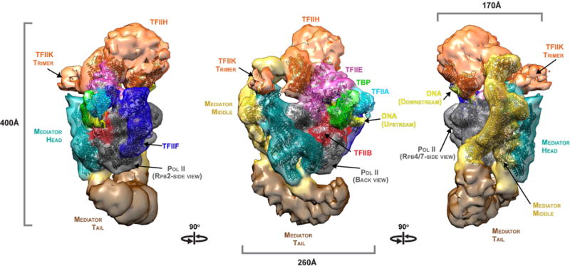
A complete, 52-protein, 2.5 million dalton, Mediator-RNA polymerase II pre-initiation complex (Med-PIC) was assembled and analyzed by cryo-electron microscopy and by chemical cross-linking and mass spectrometry. The resulting complete Med-PIC structure reveals two components of functional significance, absent from previous structures, a protein kinase complex and the Mediator-activator interaction region. It thereby shows how the kinase and its target, the C-terminal domain of the polymerase, control Med-PIC interaction and transcription.

Keywords: Mediator complex; Pre-initiation complex; RNA polymerase II carboxy-terminal domain; TFIIH; TFIIK; Transcription; cross-linking; cryo-EM; mass spectrometry.


Figures

Figure 1
Figure 1. Assembly and characterization of the Med-PIC complex
(A) Assembly pathway of the Med-PIC complex. Color scheme here and throughout: Mediator Head (aquamarine), Middle (gold), Tail (brown) as well as Pol II (grey), TFIIA (cyan), TFIIB (red), TFIIH (orange), TFIIE (magenta), TFIIF (blue) and TBP (green). (B) Glycerol gradient sedimentation of Med-PIC (upper) and PIC (lower). SDS-PAGE of gradient fractions and of Mediator, TFIIH and TBP-IIB-IIA are shown. (C) SDS-PAGE of the 52-subunit yeast Med-PIC complex (left) and the 21-subunit yeast Mediator complex for reference (right). (D) Transcripts produced from peak gradient fractions for Med-PIC complex and PIC, with and without TBP as indicated, analyzed by gel electrophoresis and autoradiography. Major transcripts are slightly larger than a 73 residue marker (position indicated on the left)
Figure 2
Figure 2. Cryo-EM structure of the Med-PIC complex
A surface representation of the cryo-EM map is shown, with views related by successive 90° rotations about a vertical axis, colored accord ing to the scheme in Figure 1A. Atomic models, computationally docked to the map as described, are shown superimposed in the same colors. See also Figures S1, S2, S4 and S6.
Figure 3
Figure 3. Cross-linking and Mass Spectrometry of the Med-PIC complex
(A) Network diagram of cross-links between subunits of the Med-PIC complex (676 of the 1642 total cross-links identified here). Subunits are colored as in Figure 1A and as indicated. Blue lines represent cross-links formed in the Med-PIC complex, including Gcn4 and TFIIS (total of 54 subunits). Green lines represent cross-links formed in a Mediator - pol II complex (33 subunits). Cross-links formed in both complexes are indicated in black. Cross-links between pol II subunits and between pol II and TFIIB, -E, -F, -S and TBP are shown in the inset. (B) Validation of cross-links. Upper panel: scoring of matches (SVM confidence score) to the sequence database (target) and to a scrambled sequence database (decoy), showing a 4% “false discovery rate” (percentage of decoy matches exceeding the acceptance threshold in this work [red line])). Lower panel: validation against near atomic structures; 397 of 1642 cross-links could be mapped to Cα-Cα distances in crystal structures of pol II (pdb:1wcm), Mediator Head module (pdb:4gwp), pol II - TFIIB (pdb:4bbr), pol II – TFIIA - TBP (pdb:1rm1), Med7C/21 and Med7N/31 dimers (pdb:1yke, pdb:3fbi). Almost all (357, or 90%) of the mappable cross-links were consistent with the 35 Å upper limit. (C) Distribution of 1642 cross-links between the 13 proteins and subcomplexes defined by the color code in panel A. The area of each bubble is proportional to the number of cross-links found between the pair of proteins and subcomplexes indicated. See also Figure S3.
Figure 4
Figure 4. Map of transcription factor – pol II interactions
Pol II is shown in standard views with its surface colored according to interactions with Mediator and GTFs. Interaction data include EM and cross-link results from this study and earlier ones (Figures 3 and S3), as well as published crystal structures.
Figure 5
Figure 5. CTD contacts drive the Mediator – pol II interaction
The binding kinetics of complete Mediator, Mediator modules, and TFIIF (analytes) to immobilized RNA polymerase II and pol II variants (surface ligands) were determined by surface plasmon resonance. (A) Single-cycle kinetic analysis of the interaction of Mediator with immobilized pol II, pol II with mutated CTD, and pol II lacking the CTD. (B) Binding of TFIIF to pol II lacking the CTD, as a control to confirm that this pol II is capable of functionally important interactions. (C) Binding of the Mediator Head module to immobilized pol II variants. (D) Binding of Mediator lacking the Tail module to immobilized pol II variants. (E) Table summarizing the data from multiple measurements of interactions between Mediator complexes and pol II variants. All kinetic data were processed using double reference subtraction (blank analyte injection and Dligand reference lane). For a subset of the data (*), binding curves were fit using a ‘Heterogeneous Ligand’ model, consistent with the availability of multiple equivalent CTD heptad repeats, otherwise a Langmuir model (1:1 binding) was used. Analyses showing no detectable binding are indicated (☥). See also Figure S5.
Figure 6
Figure 6. Position of TFIIK and CTD and stimulation of CTD phosphorylation
(A) Position of TFIIK density in the Med-PIC structure. TFIIK difference density calculated by subtraction of core Med-Pol II and PIC maps from the Med-PIC structure (Figure S7 and Materials and Methods) is shown after filtration and at the appropriate map threshold (orange density). (B) Integrative modeling reveals TFIIK subunit localizations (solid transparent density) within the Med-PIC EM map (blue mesh) that are consistent with the calculated TFIIK difference density and cross-links between TFIIK and Rad3 surface lysine residues (red spheres). (C) Model for the path of the CTD through the initiation complex. The CTD path is constrained by multiple anchor points provided by pol II and CTD-Med Head crystal structures, positioning the CTD kinase (Kin28) and cross-links obtained between a C-terminal CTD and N-terminal Med19 lysine residues (Plaschka et al., 2015; Robinson et al., 2015) (Figure 3A). (D) Close association between the Med7N-31 and Neck domain of the Head module at the Head-Middle interface forms a CTD channel that directs the CTD towards interaction with TFIIK. The stimulation of CTD phosphorylation by Mediator is explained by the role of Mediator in positioning both TFIIK and CTD for interaction. See also Figure S7.
Figure 7
Figure 7. Tail module architecture and dynamics
(A) The architectural model of free Mediator (colored subunit map) can be docked unambiguously into the Med-PIC structure (grey semi-transparent density) showing the position of the three Mediator modules and a novel connection between the Middle and Tail (dashed red oval). (B) Revised Tail module architectural map showing the position of Tail subunits and their colocalization with the Med14 C-terminus at Tail interface 1 and with Med1 at Tail interface 2. (C) Cross-links supporting the revised Tail module architecture. (D) Alternate Tail module conformations. Whereas most particles show the Tail well separated from pol II (Tail-down), a subpopulation reveal a movement of the Tail towards the upstream DNA and in close association of Tail with pol II (Tail-up). See also Figure S7.

References

    1. Allison LA, Ingles CJ. Mutations in RNA polymerase II enhance or suppress mutations in GAL4. Proceedings of the National Academy of Sciences of the United States of America. 1989;86:2794–2798. - PMC - PubMed
    1. Asturias FJ, Jiang YW, Myers LC, Gustafsson CM, Kornberg RD. Conserved structures of mediator and RNA polymerase II holoenzyme. Science. 1999;283:985–987. - PubMed
    1. Baumli S, Hoeppner S, Cramer P. A conserved mediator hinge revealed in the structure of the MED7.MED21 (Med7.Srb7) heterodimer. The Journal of biological chemistry. 2005;280:18171–18178. - PubMed
    1. Beve J, Hu GZ, Myers LC, Balciunas D, Werngren O, Hultenby K, Wibom R, Ronne H, Gustafsson CM. The structural and functional role of Med5 in the yeast Mediator tail module. The Journal of biological chemistry. 2005;280:41366–41372. - PubMed
    1. Bich C, Maedler S, Chiesa K, DeGiacomo F, Bogliotti N, Zenobi R. Reactivity and applications of new amine reactive cross-linkers for mass spectrometric detection of protein-protein complexes. Analytical chemistry. 2010;82:172–179. - PubMed

Publication types

MeSH terms