. 2016 Sep 9;12(9):e1006274.
doi: 10.1371/journal.pgen.1006274. eCollection 2016 Sep.

mSWI/SNF (BAF) Complexes Are Indispensable for the Neurogenesis and Development of Embryonic Olfactory Epithelium

Affiliations

mSWI/SNF (BAF) Complexes Are Indispensable for the Neurogenesis and Development of Embryonic Olfactory Epithelium

Christina Bachmann et al. PLoS Genet. .

Abstract

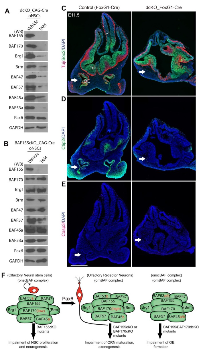
Neurogenesis is a key developmental event through which neurons are generated from neural stem/progenitor cells. Chromatin remodeling BAF (mSWI/SNF) complexes have been reported to play essential roles in the neurogenesis of the central nervous system. However, whether BAF complexes are required for neuron generation in the olfactory system is unknown. Here, we identified onscBAF and ornBAF complexes, which are specifically present in olfactory neural stem cells (oNSCs) and olfactory receptor neurons (ORNs), respectively. We demonstrated that BAF155 subunit is highly expressed in both oNSCs and ORNs, whereas high expression of BAF170 subunit is observed only in ORNs. We report that conditional deletion of BAF155, a core subunit in both onscBAF and ornBAF complexes, causes impaired proliferation of oNSCs as well as defective maturation and axonogenesis of ORNs in the developing olfactory epithelium (OE), while the high expression of BAF170 is important for maturation of ORNs. Interestingly, in the absence of BAF complexes in BAF155/BAF170 double-conditional knockout mice (dcKO), OE is not specified. Mechanistically, BAF complex is required for normal activation of Pax6-dependent transcriptional activity in stem cells/progenitors of the OE. Our findings unveil a novel mechanism mediated by the mSWI/SNF complex in OE neurogenesis and development.


Conflict of interest statement

The authors have declared that no competing interests exist.

Figures

Fig 1
Fig 1. Subunit switching in the BAF complex during differentiation of oNSCs to ORNs.
(A) Expression of BAF subunits in oNSCs and ORNs. Cell lysates were prepared from cultured oNSCs and ORNs, and blotted with BAF subunit-specific antibodies. (B) Co-immunoprecipitation of nuclear extracts from cultured oNSCs and ORNs using anti-Brg1/Brm antibodies and Western blot analyses with antibodies against BAF subunits revealed that BAF subunits integrate into Brg1/Brm-based BAF complexes. (C, D) Double-immunostaining of coronal sections of the OE from E12.5 mice revealed non-overlapping expression of oNSC-specific BAF45a/BAF53a subunits and ORN-specific BAF45b/BAF53b subunits in the developing OE. (E) A switch in subunits of BAF complexes during differentiation from oNSCs to ORNs. Abbreviations: oNSC, olfactory neural stem cell, ORN, olfactory receptor neuron; D/V, dorsal/ventral. Scale bars = 50 μm (C, D).
Fig 2
Fig 2. Expression of core BAF155 and BAF170 subunits in olfactory cell subtypes and in their mutants.
(A–D) Representative images from coronal sections of the OE at the indicated embryonic stages (D, dorsal; V, ventral). (A, B) Double-IHC analysis of the ubiquitous BAF155 subunit showed wide expression in the developing OE, including in Pax6+ oNSCs (A) and Lhx2+ ORNs (B) (yellow in overlaid images; see also S2 Fig for additional analyses). (C-G) IHC (C, D) and statistical analyses (E-G) is used to compare expression of BAF155 and BAF170 in control, in BAF155cKO_FoxG1-Cre at E10.5 (C) and in BAF170cKO_FoxG1-Cre OE at E13.5 (D). A moderate expression of BAF155 was observed in all cells types at E10.5 and E13.5 (control OE in C, D, G, arrows, BAF155moderate+ cells). BAF170 is expressed at low level at E10.5 OE and at BL and ALs of E13.5 OE (Control OE in C-F, emtry arrows, BAF170low+ cells), whereas most of cells with high expression of BAF170 were Ctip2+ neurons (control OE in D-F, filled arrows, BAF170high+ cells). Notably, loss of BAF170 causes a upregulated expression of BAF155 in Ctip2+ neurons (D, G filled arrows, BAF155high+ cells), but not in Ctip2- cells (D, G, emptry arrows). Abbreviations: D/V, dorsal/ventral; BL, basal layer; ALs, apical layers; ILs, intermediate layers. Scale bars = 25 μm (A, B, C) and 50 μm (D).
Fig 3
Fig 3. Histological analysis of the control and BAF155cKO developing OE.
(A, B) Images of sections from the nasal pit (NP; arrow) at E10.5, and in the rostral, medial and caudal OE at E11.5 immunostained for BAF155. Note that, at E11.5, the NP has invaginated into the nasal cavity in controls, but has not yet been initiated in mutants. (C, D) Images show coronal cryosections of control and BAF155cKO embryos at E13.5 and E15.5. Note the smaller OE in mutants compared with controls. (E) Quantification of OE volume and surface area at E10.5 in A (also see S1 Movie), and surface area at E13.5 and E15.5, shown as red frames in C and D, respectively. Abbreviations: NP, nasal pit; OE, olfactory epithelium; Tel, telencephalon; D/V, dorsal/ventral. Values are reported as means ± SEM (*P < 0.05, **P < 0.01, ***P < 0.001). Scale bar = 25 μm.
Fig 4
Fig 4. Depleted pool of oNSCs and diminished neurogenesis in embryonic BAF155cKO OE.
(A–F) Images of OE sections from control and BAF155cKO embryos showing IHC detection of the oNSC marker Sox2 at E10.5 (A) and in the basal layer at E13.5 (D); the SUS cell marker in apical layers at E13.5 (D); the IP marker Mash1 at E10.5 (B) and E13.5 (E); and the neuronal marker HuCD at E10.5 (C) and E13.5 (F) (see S4 Fig for additional markers). (G–L) Statistical quantification of panels A–F is shown. Note the decrease in the number of Sox2+ oNSCs at both E10 and E13.5. Compared to controls, the number of Mash1+ IPs and HuCD+ ORNs was diminished at E13.5, but not E10.5, in BAF155-deficient OE. Remarkably, the loss of BAF155 did not affect the genesis of Sox2+ SUS cells at E13.5. Abbreviations: D/V, dorsal/ventral; BL, basal layer; ALs, apical layers; ILs, intermediate layers. Values are reported as means ± SEM (*P < 0.05, **P < 0.01, ***P < 0.001; NS, not significant). Scale bars = 25 μm (A–C) and 50 μm (D–F).
Fig 5
Fig 5. Cell cycle indexes of oNSCs in the developing OE show selective defects in BAF155cKO mutants.
(A) Images of double-IHC for either the oNSC marker Pax6 or the IP marker Mash1 and the marker of mitotically active cells, pHH3, in E10.5 OE. (C, D) Statistical analyses showed that the loss of BAF155 led to a selective decrease in pHH3+ oNSCs at early stages (E10.5–E11.5) (B), Pax6+ oNSCs at 10.5 (C), and basal pHH3+ oNSCs at E13.5 (B), but not apical pHH3+ SUS cells (B) or pHH3+/Tbr2+ IPs (D). (E, F) Experimental paradigm in which mutant and control embryos were labeled with CIdU for 24 hours (to mark cells both in and exiting from the cell cycle) and IdU for 1 hour (to label progenitors in S-phase) by injection of the corresponding nucleoside analogs. (G–J) Images showing triple IHC at E13.5 for CIdU (red), IdU (magenta) and proliferation marker Ki67 (green) in control and BAF155cKO mutants. Middle panels: higher magnifications images of areas indicated by white boxes (G). (K, L) Statistical analyses of proliferation indexes (i.e., the number of IdU+/Ki67+ cells per total number of Ki67+ cells) for cells in panel I, showing a significantly lower value in mutant OE at the basal side, but not at the apical side (K). No significant differences were found in the exit index (i.e., number of CidU+/Ki67- cells per total number of CidU+ cells) for cells in panel J between controls and mutants (L). Values are reported as means ± SEM (*P < 0.05, **P < 0.01, ***P < 0.001; NS, not significant). Scale bars = 25 μm (A) and 50 μm (G–J).
Fig 6
Fig 6. BAF155 and Pax6 synergistically control neurogenesis in embryonic OE.
(A) Immunoprecipitation of BAF155 from cultured OE NSCs and Western blot analysis using an anti-Pax6 antibody demonstrated that BAF155 interacts with Pax6 in OE cells. (B–D) Cultured OE NSCs from control or BAF155cKO or dcKO embryos were electroporated with pCON/P3 (see B), a Pax6-dependent reporter construct, and an EGFP expression plasmid. After 2 days of in vitro culture (DIV), the electroporated cells were collected and analyzed by Western blotting with the indicated antibodies. Western blotting (C) and statistical analyses (D) indicated that compared to cultured OE cell control, the Pax6-dependent transcriptional activity was moderate (in BAF155cKO mutants) and severely diminished in dcKO mutants. (E–G) Images show IHC detection of the oNSC marker Sox2 (E), the neuronal progenitor marker Mash1 (F), and the post-mitotic neuron marker HuCD (G) in OE sections from the indicated transgenic embryos. (H–J) Statistical comparisons indicated increased decreased numbers of progenitors and neurons in the OE of BAF155cKO mice compared with control mice. These phenotypic features of BAf155cKO (BAF155fl/fl_Pax6+/+_FoxG1-Cre) mice were dramatically exacerbated by additional heterozygous loss of one Pax6 allele (BAF155fl/fl_Pax6fl/+_FoxG1-Cre). Abbreviations: D/V, dorsal/ventral. Values are reported as means ± SEM (*P < 0.05, **P < 0.01, ***P < 0.001; NS, not significant). Scale bars = 25 μm (E) and 50 μm (F, J).
Fig 7
Fig 7. Absence of mature OSNs and their axonal tracts in BAF155-deficient mutants.
(A) Coronal sections of the OE and frontal telencephalon of E13.5 and E15.5 embryos (S5C Fig) control and BAF155-deficient embryos were analyzed by IHC using anti-Tuj and anti-NCAM antibodies (S5C Fig). The axons of immature OSNs (Tul+NCAM+) reached the OB in control embryos, but failed to grow out and generate a fibro-cellular mass in BAF155-deficient embryos. (B) IHC analyses of cross sections through the head at E15.5 using an anti-OMP antibody, which labels mature OSNs and their axons, revealed a severely diminished number of OMP+ mature OSNs in the OE and a lack of axonal connectivity between the OE and rostral cortex in BAF155cKO mice compared with control mice (indicated by arrows). (C) Statistical analysis of OMP+ mature ORNs in control and BAF155cKO OE at E13.5 and E15.5. (D) Tracing the olfactory nerve with DiI at E17.5. Bright-field and fluorescent images are shown in upper and lower panels, respectively. In controls, the olfactory nerve and axons projecting to the OB are clearly outlined by DiI. In addition, these OB neurons, which have taken up DiI and project their exons via the LOT to the primary olfactory cortex, were also seen in control embryos (indicated by arrows), whereas no DiI-positive axonal projection pattern from the OE to the OB toward the olfactory cortex was detectable in the mutants. Abbreviations: ON, olfactory nerve, LOT, lateral olfactory tract. Values are reported as means ± SEM (***P < 0.001). Scale bars = 100 μm (A) and 150 μm (B).
Fig 8
Fig 8. High expression of BAF170 is important for maturation of ORNs.
(A, C, E, G, I, K, M) Images of IHC for markers: Sox2 (oNSCs, A), pHH3 (M-phase marker, C), Mash1 (IPs, E), HuCD, Lhx2 (immature ORNs, G and I respectively), Ctip2 (maturing ORNs, K) and OMP (mature ORNs, M) with OE sections from control and BAF170cKO embryos at E13.5 (A, C, E, G, I, K) and E15.5 (M). (B, D, F, H, J, L, N) Statistical quantification of panels (A, C, E, G, I, K, M) is shown. Compared to control, loss of BAF170 in BAF170cKO OE led to diminished number of Ctip2+OMP+ mature ORNs, but not pHH3+Sox2+ proliferating cells or Mash1+ IPs or HuCD+Lhx2+ immature ORNs. Abbreviations: D/V, dorsal/ventral; BL, basal layer; ALs, apical layers; ILs, intermediate layers. Values are reported as means ± SEM (*P < 0.05, **P < 0.01, ***P < 0.001; NS, not significant). Scale bar = 50 μm.
Fig 9
Fig 9. BAF155 and BAF170 expression are indispensable for OE development.
(A, B) Expression of BAF subunits in dcKO (A) and BAF155cKO (B) cultured oNSCs was shown. (A) WB analyses revealed after 3 days of TAM treatment, expression of all known onscBAF subunits is nearly-completely lost in dcKO_CAG-Cre cultured oNSCs (A), whereas expression of most onscBAF subunits (except BAF57) is preserved in BAF155-deficient oNSCs (B). (C, D) IHC analyses using antibodies that specifically label oNSCs, SUS cells (Sox2 in C), immature ORNs (Tuj in C), and maturing ORNs (Ctip2 in D). BAF155/BAF170dcKO_FoxG1-Cre mutants lack OE at E11.5 (white arrows). (E) IHC analyses revealed no detectable Casp3+ apoptotic cells in both dcKO and control OE. (F) Schema shows the BAF subunits of BAF complexes identified in oNSCs and ORNs, as well as a summary of BAF mutant phenotypes in OE development. Abbreviations: OE, olfactory epithelium; Tel, telencephalon; Di, diencephalon. Scale bar = 500 μm.

References

    1. Treloar HB, Miller AM, Ray A, Greer CA (2010) Development of the Olfactory System. In: Menini A, editor. The Neurobiology of Olfaction. Boca Raton (FL). - PubMed
    1. Suzuki J, Osumi N (2015) Neural crest and placode contributions to olfactory development. Current topics in developmental biology 111: 351–374. 10.1016/bs.ctdb.2014.11.010 - DOI - PubMed
    1. Buck LB (2005) Unraveling the sense of smell (Nobel lecture). Angewandte Chemie 44: 6128–6140. - PubMed
    1. Cau E, Casarosa S, Guillemot F (2002) Mash1 and Ngn1 control distinct steps of determination and differentiation in the olfactory sensory neuron lineage. Development 129: 1871–1880. - PubMed
    1. Cau E, Gradwohl G, Fode C, Guillemot F (1997) Mash1 activates a cascade of bHLH regulators in olfactory neuron progenitors. Development 124: 1611–1621. - PubMed

LinkOut - more resources