. 2016 Sep 9;11(9):e0161909.
doi: 10.1371/journal.pone.0161909. eCollection 2016.

In Silico and Biochemical Characterization of Lysozyme-Like Proteins in the Rat

Affiliations

In Silico and Biochemical Characterization of Lysozyme-Like Proteins in the Rat

Ganapathy Narmadha et al. PLoS One. .

Abstract

Background: Spermatogenesis and sperm maturation in the male reproductive tract is dictated by a variety of proteins secreted in the testis and epididymis. Though the proteome of these tissues is known, the functional role of many of these proteins remains uncharacterized. In this study, we characterize the rat Lysozyme-like (Lyzl) genes and proteins.

Methods: In silico tools were used to predict the primary, secondary and tertiary structures. Reverse transcription PCR, immunofluorescence and immunoblotting were used to determine the expression pattern. Lysozyme like enzyme activity was assessed by standard assays.

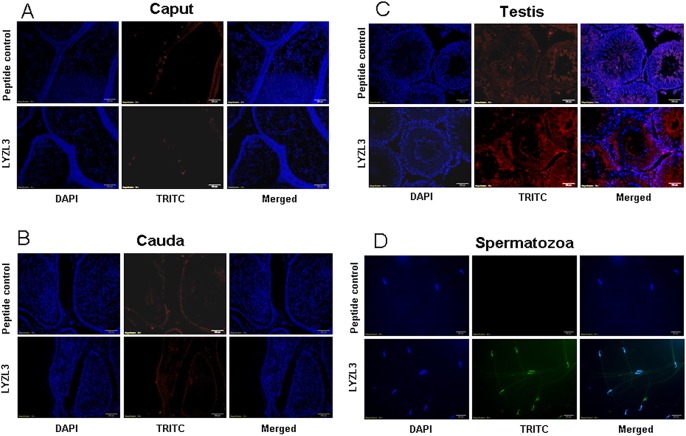
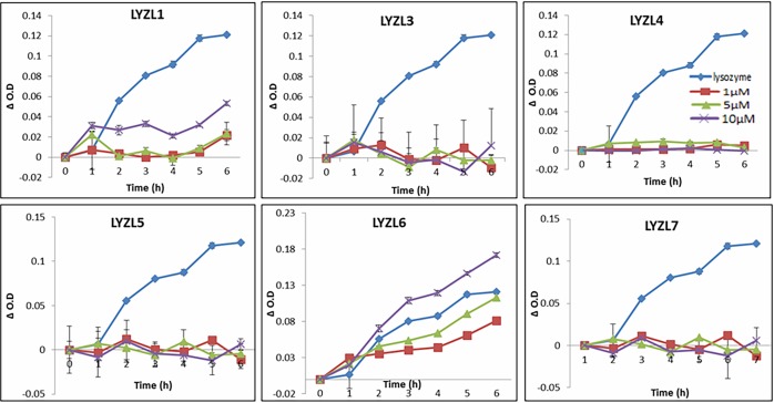
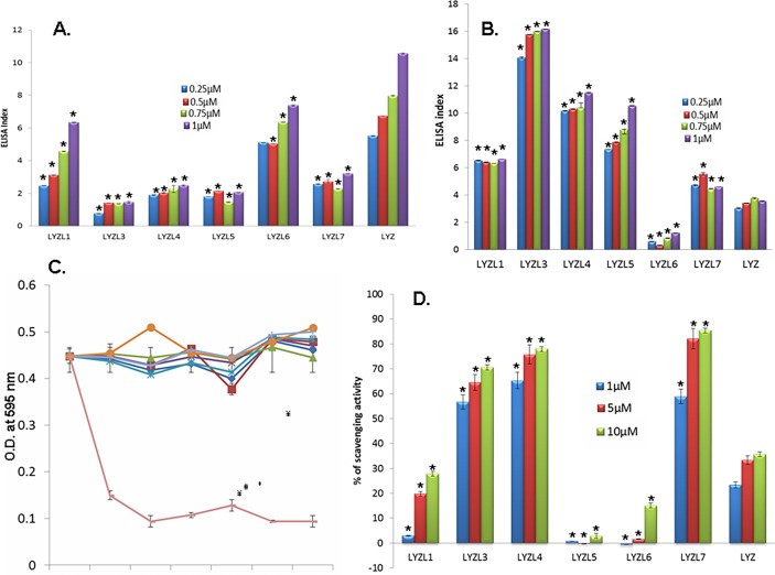
Results: Six rat Lyzl genes namely Lyzl1, Lyzl3, Lyzl4, Lyzl5, Lyzl6 and Lyzl7 were found to be highly conserved among the vertebrates with higher homology to mouse counterparts than with human counterparts. All the LYZL proteins contained the characteristic 4 disulfide bridges similar to c-type lysozyme. Only LYZL 1 and 6, conserved the active site amino acids of the lysozyme. Molecular modeling studies indicated that LYZL proteins exhibit strikingly similar three-dimensional structures among themselves. The secondary structure analysis of the recombinant LYZL proteins indicated the presence of α-helix, β-sheet and random coil with α-helix being the majority. Docking studies indicated the peptidoglycan binding nature of LYZL proteins. All the rat Lyzl mRNA transcripts (Lyzl1, Lyzl3, Lyzl4, Lyzl5, Lyzl6 and Lyzl7) are predominantly expressed in testes though some of them are expressed in tissues other than reproductive tract. Their expression was androgen independent. The rat LYZL proteins are localized in the germinal epithelium and on the spermatozoa. Recombinant LYZL1 and 6 possessed muramidase, isopeptidase and antibacterial activities. The mechanism of antibacterial action of LYZL1 and LYZL6 involved bacterial membrane damage and leakage of cellular contents. Only LYZL1 and 6 possess peptidoglycan binding ability, whereas LYZL3, LYZL4 and LYZL5 possess hyaluronan binding ability suggesting a possible functional divergence of these proteins. LYZL3, LYZL4 and LYZL7 possessed free radical scavenging property, suggesting that they may act as antioxidants.

Conclusion: The divergent properties of LYZL proteins indicate that they may have a role in sperm function, innate immunity and other physiological process as well.


Conflict of interest statement

The authors have declared that no competing interests exist.

Figures

Fig 1
Fig 1. Alignment of rat lysozyme and LYZL proteins.
Green color boxed amino acids represent the active site and black arrows indicate the conserved 8 cysteines. Sequence alignment of some rat LYZLs was reported by us earlier [24] and this figure presents a comprehensive analysis of all rat LYZLs.
Fig 2
Fig 2. Genomic neighbourhood analysis of rat, mouse and human Lyzl genes.
Green arrows with names represent the genes and directions of the arrow represent the direction of sense strand. Black, red and green arrows indicate noncoding, pseudo and hypothetical genes respectively. Numbers in the beginning of each row indicates the chromosome number and are shown in the order of rat, mouse and human.
Fig 3
Fig 3. Circular dichroism spectra of recombinant LYZL proteins measured in terms of mean residue ellipticity (MRE).
Fig 4
Fig 4. Modelled structure of LYZL proteins showing β- sheets in yellow, helices in red, loop regions in green and disulphide bonds in spheres.
Fig 5
Fig 5. Molecular modeling showing the superimposition of rat LYZL and NAG complex with chicken lysozyme-NAG complex.
NAG is represented in the form of stick model and the protein in cartoon model.
Fig 6
Fig 6. Expression of Lyzl genes in the male reproductive and non- reproductive tissues.
RNA isolated from different tissues of rats were reverse transcribed and used for gene specific PCR. Gapdh was used as internal control. Cp-caput, Co-corpus, Cd-cauda, T- testes, Sv-seminal vesicle, P-prostate, B-Brain, H- Heart, Lu-Lungs, Li-Liver, K-Kidney, Sp-Spleen, O-Ovary, Ut-Uterus, Ce-Cervix, Gapdh-Glyceraldehyde 3 phosphate. Expression pattern of some rat LYZL genes were reported by us earlier [24]and this figure includes our previously reported data to present a comprehensive analysis of all rat LYZL genes.
Fig 7
Fig 7. Developmental regulation of Lyzl genes in epididymidis and testes.
RT PCR for Lyzl genes in epididymis and testes collected from rats of different age groups. 10, 20, 30, 40, 50 and 60 are the age of rats in days. cp- caput, cd-cauda. Gapdh was used as the internal control. Expression pattern of some rat LYZL genes were reported by us earlier [24] and this figure includes our previous data to present a comprehensive analysis of all rat LYZL genes.
Fig 8
Fig 8. LYZL protein expression in male reproductive tract.
Immunoblotting of LYZL proteins in male reproductive tract tissues. The immunoblots were probed with immune serum specific to each protein. Cp-caput, Cd-cauda, T-testes, Sv-seminal vesicles, P-prostate, Spm-sperm, Rec-recombinant protein.
Fig 9
Fig 9. Immunolocalization of LYZL1.
Serial sections of the rat testes and epididymides were incubated with antigen preadsorbed immune serum (peptide control) or immune serum raised against LYZL1, followed by TRITC (Tetramethylrhodamine-5-(and-6)-Isothiocyanate (5(6)) tagged secondary antibody and counter stained with DAPI (4',6-diamidino-2-phenylindole) nuclear stain. Spermatozoa were stained with FITC (fluorescein Isothiocyanate) tagged secondary antibody.
Fig 10
Fig 10. Immunolocalization of LYZL3.
Serial sections of the rat testes and epididymides were incubated with antigen preadsorbed immune serum (peptide control) or immune serum raised against LYZL3, followed by TRITC (Tetramethylrhodamine-5-(and-6)-Isothiocyanate (5(6)) tagged secondary antibody and counter stained with DAPI (4',6-diamidino-2-phenylindole) nuclear stain. Spermatozoa were stained with FITC (fluorescein Isothiocyanate) tagged secondary antibody.
Fig 11
Fig 11. Immunolocalization of LYZL4.
Serial sections of the rat testes and epididymides were incubated with antigen preadsorbed immune serum (peptide control) or immune serum raised against LYZL4, followed by TRITC (Tetramethylrhodamine-5-(and-6)-Isothiocyanate (5(6)) tagged secondary antibody and counter stained with DAPI (4',6-diamidino-2-phenylindole) nuclear stain. Spermatozoa were stained with FITC (fluorescein Isothiocyanate) tagged secondary antibody. Panel D showing LYZL4 localization was reported by us earlier [24] and the same figure is being used for a comprehensive presentation of results.
Fig 12
Fig 12. Immunolocalization of LYZL5.
Serial sections of the rat testes and epididymides were incubated with antigen preadsorbed immune serum (peptide control) or immune serum raised against LYZL5, followed by TRITC (Tetramethylrhodamine-5-(and-6)-Isothiocyanate (5(6)) tagged secondary antibody and counter stained with DAPI (4',6-diamidino-2-phenylindole) nuclear stain. Spermatozoa were stained with FITC (fluorescein Isothiocyanate) tagged secondary antibody.
Fig 13
Fig 13. Immunolocalization of LYZL6.
Serial sections of the rat testes and epididymides were incubated with antigen preadsorbed immune serum (peptide control) or immune serum raised against LYZL6, followed by TRITC (Tetramethylrhodamine-5-(and-6)-Isothiocyanate (5(6)) tagged secondary antibody and counter stained with DAPI (4',6-diamidino-2-phenylindole) nuclear stain. Spermatozoa were stained with FITC (fluorescein Isothiocyanate) tagged secondary antibody.
Fig 14
Fig 14. Immunolocalization of LYZL7.
Serial sections of the rat testes and epididymides were incubated with antigen preadsorbed immune serum (peptide control) or immune serum raised against LYZL7, followed by TRITC (Tetramethylrhodamine-5-(and-6)-Isothiocyanate (5(6)) tagged secondary antibody and counter stained with DAPI (4',6-diamidino-2-phenylindole) nuclear stain. Spermatozoa were stained with FITC (fluorescein Isothiocyanate) tagged secondary antibody.
Fig 15
Fig 15. Muramidase activity of LYZL proteins.
1 μM (■), 5 μM (▲) and 10 μM (X) of recombinant LYZL proteins were incubated with 2 ml of M. lysodeikticus culture and the O.D monitored at 450nm. Chicken lysozyme (1μM; ♦) was used as a positive control. Values shown are mean ± SD.
Fig 16
Fig 16. Isopeptidase activity of recombinant LYZL proteins.
1 μM (■), 5 μM (▲) and 10 μM (X) of recombinant LYZL proteins were incubated with 1.75 mM L-γ-Glu-pNA in 0.05 M 3-(N-morpholino) propane sulfonic acid (MOPS) buffer, pH 7, containing 0.01M NaCl and the O.D monitored at 405nm. Chicken lysozyme (1μM; ♦) was used as positive control. Values shown are mean ± SD. The isopeptidase activity of LYZL4 was reported by us earlier [24] and the same data is included in this figure for a comprehensive presentation of results.
Fig 17
Fig 17. Antibacterial activity of LYZL proteins.
E. coli XL-1 blue grown to mid-log phase were incubated with 0 (♦), 10 (■), 25 (▲), 50 (X) and 100 (*) μg/ml of recombinant protein for 30, 60, 90 and 120 min after the start of incubation. The assay mixtures were diluted, plated on LB agar plates and the colonies hand counted. Values shown are Mean ± S.D. The isopeptidase activity of LYZL4 was reported by us earlier [24] and the same data is included in this figure for a comprehensive presentation of results.
Fig 18
Fig 18. Membrane permeabilizing ability of LYZL proteins.
(A) Dye based membrane potential measurement in E. coli treated with recombinant LYZL proteins using flow cytometry. A- MFI measurements of the cells in FITC, PE-Texas Red A and PerCP-Cy5-5-A channels. (B) Membrane potential measured in terms of ratio of mean fluorescence intensity of red/green. (C) Effect of recombinant LYZL1 and LYZL6 on the morphology of E. coli. Scanning electron micrographs of E. coli treated with 100 μg/ml recombinant LYZL proteins for 2 h. Chicken Lysozyme (10 μg/ml) was used as positive control.
Fig 19
Fig 19. Substrate binding ability of LYZL proteins.
(A) ELISA based peptidoglycan binding assay. 40 μg/ml peptidoglycan coated plate was incubated with 0.25, 0.5, 0.75 and 1μM of the recombinant proteins. Affinity of protein bound to peptidoglycan was measured in terms of color produced during development after probing with corresponding primary and secondary antibody. (B) ELISA based hyaluronan binding assay. 40 μg/ml hyaluronan coated into the wells of a microtitre plate was incubated with 0.25, 0.5, 0.75 and 1μM of the recombinant protein. Protein binding to peptidoglycan was measured by ELISA based colour detection. ELISA index was calculated by subtracting the average O.D of negative control and dividing the resultant by the negative control O.D. (C) Hyaluronidase activity of LYZL proteins. Hyaluronan mixed agarose incubated with different concentrations of recombinant LYZL proteins. The amount of cetyl pyridinium chloride precipitate cleared is a measure of hyaluronidase activity. Hyaluronidase was used as positive control. (D) Free radical scavenging assay. DPPH solution was incubated with varying concentrations of recombinant LYZL protein and the decrease in O.D was measured at 517 nm. Lysozyme was used a control. Radical scavenging activity was expressed in terms of percentage. Values shown are mean ± SD. * indicates p<0.05 compared to the corresponding concentration of lysozyme.

References

    1. Li J, Liu F, Liu X, Liu J, Zhu P, Wan F, et al. (2011) Mapping of the Human Testicular Proteome and its Relationship With That of the Epididymis and Spermatozoa. molecular and cellular proteomics 3: 1–11. - PMC - PubMed
    1. Zhou Y, Zheng M, Shi Q, Zhang L, Zhen W, Chen W, et al. (2008) An epididymis-specific secretory protein HongrES1 critically regulates sperm capacitation and male fertility. PloS one 3: e4106–e4106. 10.1371/journal.pone.0004106 - DOI - PMC - PubMed
    1. Kirchhoff C (1998) Molecular characterization of epididymal proteins. Reviews of reproduction 3: 86–95. - PubMed
    1. Jiborn T, Abrahamson M, Wallin H, Malm J, Lundwall A, Gadaleanu V, et al. (2004) Cystatin C is highly expressed in the human male reproductive system. Journal of andrology 25: 564–572. - PubMed
    1. Jin YZ, Bannai S, Dacheux F, Dacheux JL, Okamura N (1997) Direct evidence for the secretion of lactoferrin and its binding to sperm in the porcine epididymis. Molecular reproduction and development 47: 490–496. - PubMed

LinkOut - more resources