. 2016 Oct 25;7(43):70404-70419.
doi: 10.18632/oncotarget.11879.

Androgen receptor differentially regulates the proliferation of prostatic epithelial cells in vitro and in vivo

Affiliations

Androgen receptor differentially regulates the proliferation of prostatic epithelial cells in vitro and in vivo

Shu Yang et al. Oncotarget. .

Abstract

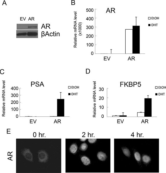
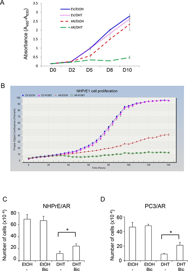
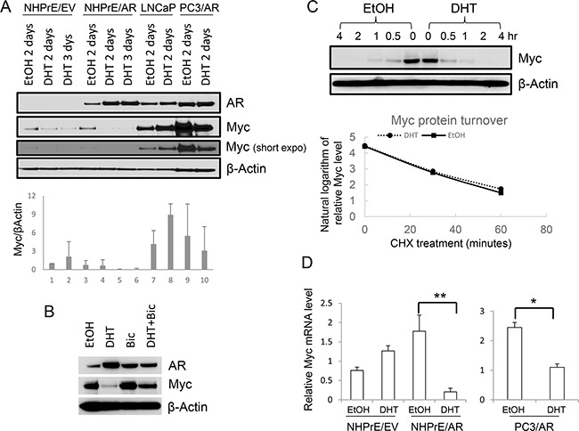
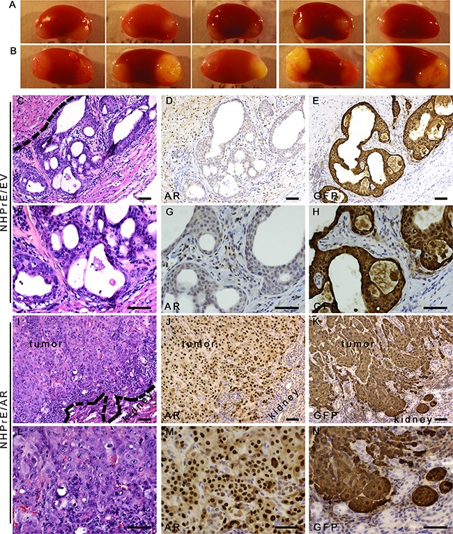
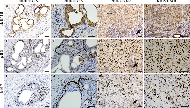
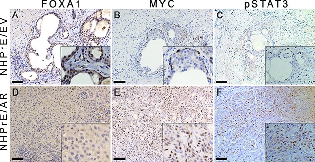
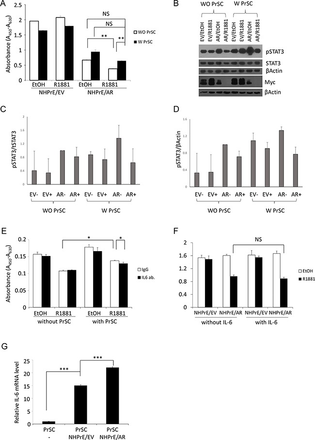
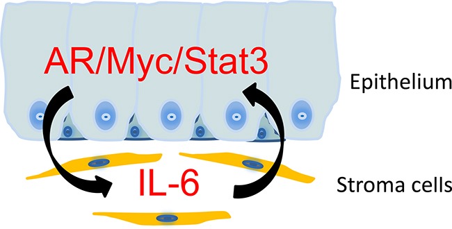
Androgens regulate the proliferation and differentiation of prostatic epithelial cells, including prostate cancer (PCa) cells in a context-dependent manner. Androgens and androgen receptor (AR) do not invariably promote cell proliferation; in the normal adult, endogenous stromal and epithelial AR activation maintains differentiation and inhibits organ growth. In the current study, we report that activation of AR differentially regulates the proliferation of human prostate epithelial progenitor cells, NHPrE1, in vitro and in vivo. Inducing AR signaling in NHPrE1 cells suppressed cell proliferation in vitro, concomitant with a reduction in MYC expression. However, ectopic expression of AR in vivo stimulated cell proliferation and induced development of invasive PCa in tissue recombinants consisting of NHPrE1/AR cells and rat urogenital mesenchymal (UGM) cells, engrafted under renal capsule of adult male athymic mice. Expression of MYC increased in the NHPrE1/AR recombinant tissues, in contrast to the reduction seen in vitro. The inhibitory effect of AR signaling on cell proliferation in vitro were reduced by co-culturing NHPrE1/AR epithelial cells with prostatic stromal cells. In conclusion, these studies revealed that AR signaling differentially regulates proliferation of human prostatic epithelia cells in vitro and in vivo through mechanisms involving stromal/epithelial interactions.

Keywords: androgen receptor; prostate.


Conflict of interest statement

CONFLICTS OF INTEREST

The authors declare no conflict of interest.

Figures

Figure 1
Figure 1. Ectopic expression of AR conferred functional AR-mediated androgen signaling in NHPrE1 cells
Retroviral vector pLNCX or pLNCX-AR was used to generate NHPrE1 cells with empty vector (EV) control or AR transgene. A. Western blot to analyze the expression of AR in NHPrE1/EV (EV) or NHPrE1/AR (AR) cells. Beta-actin served as a loading control. B-D. quantitative (q)RT-PCR to assess the levels of AR (B) and androgen responsive genes PSA (C) and FKBP5 (D). Androgen treatment (DHT, 10 nM) induced the expression of PSA and FKBP5. The expression of GAPDH was used to normalize the qPCRs. E. immunofluorescence staining of AR. NHPrE1/AR cells were cultured in androgen-depleted medium for 24 hours and then treated with R1881 (1nM) for 2 or 4 hours. Immunofluorescence staining of AR was conducted to examine the nuclear translocation of AR upon androgen treatment.
Figure 2
Figure 2. Androgen differentially regulated prostatic cell proliferation
The proliferation of NHPrE1 cells with or without AR expression was assessed by using both WST-1 A. and IncuCyte B. methods. NHPrE1/EV cells and NHPrE1/AR cells were cultured in the absence or presence of androgens (10 nM DHT or 1 nM R1881). Androgen treatment had negligible effects on the proliferation of empty vector control cells, but suppressed proliferation of AR-expressing NHPrE1 cells (panel B, p<0.01 from 36 hour onward, comparison between ethanol- and R1881- treated NHPrE1/AR cells). Overall, compared with NHPrE1/EV cells, NHPrE1/AR cells displayed suppressed cell proliferation. C and D, blocking AR attenuated androgen-induced proliferation inhibition. NHPrE1/AR (C) or PC3/AR cells (D) were cultured with or without androgen (10 nM DHT) in the presence or absence of 10 μM bicalutamide (Bic) for 5 days. While DHT suppressed the proliferation of NHPrE1/AR and PC3/AR cells, addition of bicalutamide attenuated this inhibitory effect of androgens. *p<0.05, t-test.
Figure 3
Figure 3. Androgen differentially regulated MYC expression
A. Western blot for AR and MYC in prostatic cells. Androgen (DHT, 10 nM) treatment resulted in up-regulation of MYC in LNCaP cells, but down-regulation of MYC in NHPrE1/AR and PC3/AR cells. Lower panel is the quantification of MYC Western blot. B. Western blot for AR and MYC. NHPrE1/AR cells were cultured in androgen-depleted medium for 2 days with or without the addition of 10 nM DHT and/or 10 μM Bicalutamide (Bic). Bicalutamide treatment reversed androgen-mediated reduction of MYC. C. Analysis of MYC protein stability. Cycloheximide chase analyses were conducted using NHPrE1/AR cells to determine whether androgen treatment affected the turnover of MYC. NHPrE1/AR cells were treated with 50 μg/ml cycloheximide to block protein synthesis in the presence or absence of 10 nM DHT and harvested at different time points post treatment. Androgen treatment did not alter the stability of MYC protein in NHPrE1/AR cells. Lower panel is the semi-logarithm plot of MYC levels at different times of cycloheximide treatment. D. qRT-PCR to assess the levels of MYC mRNA in NHPrE1/EV, NHPrE1/AR, and PC3/AR cells. The expression of GAPDH was used to normalize the qPCRs. DHT treatment significantly decreased the level of MYC mRNA in NHPrE1/AR and PC3/AR cells. *p<0.05, **p<0.01, t-test.
Figure 4
Figure 4. Ectopic-expression of AR transformed NHPrE1 cells in vivo
NHPrE1/EV or NHPrE1/AR cells were recombined with rat UGM and grafted in vivo. A and B. gross morphology of renal subcapsular grafts. A, grafts derived from empty vector control NHPrE1/EV cells showed limited growth; B, grafts derived from NHPrE1/AR cells grew extensively. C-N. H&E and IHC staining performed on serial sections derived from NHPrE1/EV (C-H) or NHPrE1/AR (I-N) grafts. F-H and L-N are higher magnification pictures of C-E and I-K, respectively. Broken lines in panels C and I indicate the interface between the NHPrE1 grafts and host kidneys. While a clear boundary existed between the NHPrE1/EV graft and host kidney (C), NHPrE1/AR tumors focally invaded renal parenchyma (I-K). While epithelial cells in NHPrE1/AR grafts were positive for AR by IHC staining (J and M), epithelial cells in NHPrE1/EV graft showed little AR immunoreactivity (D and G). Stromal cells in NHPrE1/EV grafts (derived from rat UGM) were positive for AR staining (D and G). Epithelial cells in NHPrE1/EV grafts showed positive IHC staining for GFP and formed glandular structures (E and H); whereas GFP-tagged NHPrE1/AR cells (K and N) formed invasive carcinomas. Scale bars represent 25 μm.
Figure 5
Figure 5. Histology of NHPrE1/EV and NHPrE1/AR grafts
IHC stains for cytokeratin 8/18 (ck8/18, luminal epithelial cell marker), p63 (basal epithelial cell marker), and ki67 (cell proliferation marker) were performed on serial sections derived from NHPrE1/EV A, B, E, F, I, and J. or NHPrE1/AR grafts C, D, G, H, K, and L. B, D, F, H, J, and L are higher magnification photomicrographs of A, C, E, G, I, and K, respectively. Arrows in panels C, G, and K indicate host kidney. While NHPrE1/EV control cells formed glandular structures consisting of cytokeratin 8/18-positive luminal epithelial cells (A and B) and p63-positive basal cells (E and F), NHPrE1/AR cells formed invasive carcinomas that were positive for both cytokeratin 8/18 (C and D) and p63 IHC staining (G and H). Ki67 was barely detectable in luminal epithelial cells from NHPrE1/EV grafts (I and J), but was present in many basal cell nuclei in NHPrE1/EV grafts (I and J), as well as in malignant cells in NHPrE1/AR grafts (K and L). Scale bars represent 25 μm.
Figure 6
Figure 6. Expression of MYC and pSTAT3, but not FOXA1 was increases in NHPrE1/AR grafts
IHC staining for FOXA1, MYC, and pSTAT3 was performed on serial sections derived from NHPrE1/EV and NHPrE1/AR grafts. Insets in each panel are higher magnification photomicrographs. While NHPrE1/EV grafts showed weak IHC staining for FOXA1 A. FOXA1 was not expressed in the majority of epithelial cells in NHPrE1/AR grafts D. While only a few basal cells in NHPrE/EV grafts displayed immunoreactivity MYC B. MYC was highly expressed in tumor cells from NHPrE1/AR grafts E. Malignant cells in some areas of NHPrE1/AR grafts were also positive for pSTAT3 F. whereas pSTAT3 levels were negligible in epithelial cells of NHPrE1/EV grafts C.
Figure 7
Figure 7. Stromal/epithelial interactions are involved in modulating the proliferation of NHPrE1 cells
A. WST-1 cell proliferation assay to assess proliferation of NHPrE1 cells in the presence or absence of prostatic stromal cells (PrSC). NHPrE1/EV and NHPrE1/AR cells were co-cultured with PrSC for 5 days. While androgen treatment (1 nM R1881) inhibited proliferation of NHPrE1/AR cells in the absence of PrSC, co-culture with PrSC stimulated proliferation of NHPrE1/AR cells and partially reversed the proliferation inhibitory effect of androgens seen in vitro. ** p<0.01, t-test. B. Western blots to assess levels of pSTAT3 in NHPrE1/EV or NHPrE1/AR cells cultured with or without PrSC for 2 days. Levels of pSTAT3 (Tyr-705) and total STAT3 in NHPrE1/EV and NHPrE1/AR cells were compared; beta-Actin served as loading control. Co-culture with PrSC increased the levels of pSTAT3 but not MYC in NHPrE1 cells. C and D. quantification of pSTAT3 Western blot. The levels of pSTAT3 were normalized by total STAT3 (C) or by beta-Actin (D). E. blocking IL-6 attenuated the stimulatory effect of PrSC on NHPrE1 cell proliferation. NHPrE1/AR were cultured with or without PrSC cells in the presence or absence of IL-6 neutralizing antibody for 3 days. Anti-IL-6 attenuated the proliferation stimulation effect of PrSC. * p<0.05, t-test. Similar trend was observed in additional independent experiments. F. WST-1 cell proliferation assay. NHPrE1 cells were cultured in the presence or absence of androgens (1 nM R1881) with or without the addition of IL-6 (25 ng/ml). Addition of IL-6 to the cell culture medium did not induce the proliferation of NHPrE1 cells. G. RT-qPCR to assess expression of IL-6 in PrSC cells. Prostate stromal cells (PrSC) were cultured in the presence or absence of NHPrE1/EV (N/EV) or NHPrE1/AR (N/AR) cells. Co-culture with prostate epithelial cells stimulated production of IL-6 mRNA in PrSC cells. ***p<0.001, t-test.
Figure 8
Figure 8. A schematic representation on the stromal/epithelial interaction in NHPrE1/AR tissue recombinants
NHPrE1 cells produce factors that induce the expression of IL-6 in prostate stroma. Prostate stromal cells promote the proliferation of NHPrE1 cells via a mechanism that involves the induction of MYC and pSTAT3 in NHPrE1/AR cells. The combined expression of AR, MYC, and pSTAT3 may transform NHPrE1 cells in vivo.

References

    1. Huggins C. Effect of Orchiectomy and Irradiation on Cancer of the Prostate. Annals of surgery. 1942;115:1192–200. - PMC - PubMed
    1. Debes JD, Tindall DJ. Mechanisms of androgen-refractory prostate cancer. N Engl J Med. 2004;351:1488–90. - PubMed
    1. Wang Y, Hayward S, Cao M, Thayer K, Cunha G. Cell differentiation lineage in the prostate. Differentiation; research in biological diversity. 2001;68:270–9. - PubMed
    1. Shabsigh A, Chang DT, Heitjan DF, Kiss A, Olsson CA, Puchner PJ, Buttyan R. Rapid reduction in blood flow to the rat ventral prostate gland after castration: preliminary evidence that androgens influence prostate size by regulating blood flow to the prostate gland and prostatic endothelial cell survival. Prostate. 1998;36:201–6. - PubMed
    1. Shabsigh A, Tanji N, D'Agati V, Burchardt T, Burchardt M, Hayek O, Shabsigh R, Buttyan R. Vascular anatomy of the rat ventral prostate. Anat Rec. 1999;256:403–11. - PubMed