. 2016 Sep 27;7(39):64203-64220.
doi: 10.18632/oncotarget.11887.

F5-peptide induces aspermatogenesis by disrupting organization of actin- and microtubule-based cytoskeletons in the testis

Affiliations

F5-peptide induces aspermatogenesis by disrupting organization of actin- and microtubule-based cytoskeletons in the testis

Ying Gao et al. Oncotarget. .

Abstract

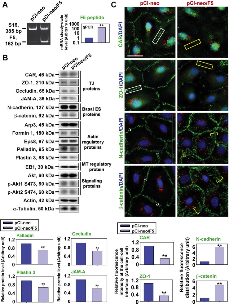
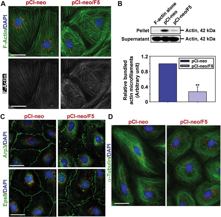
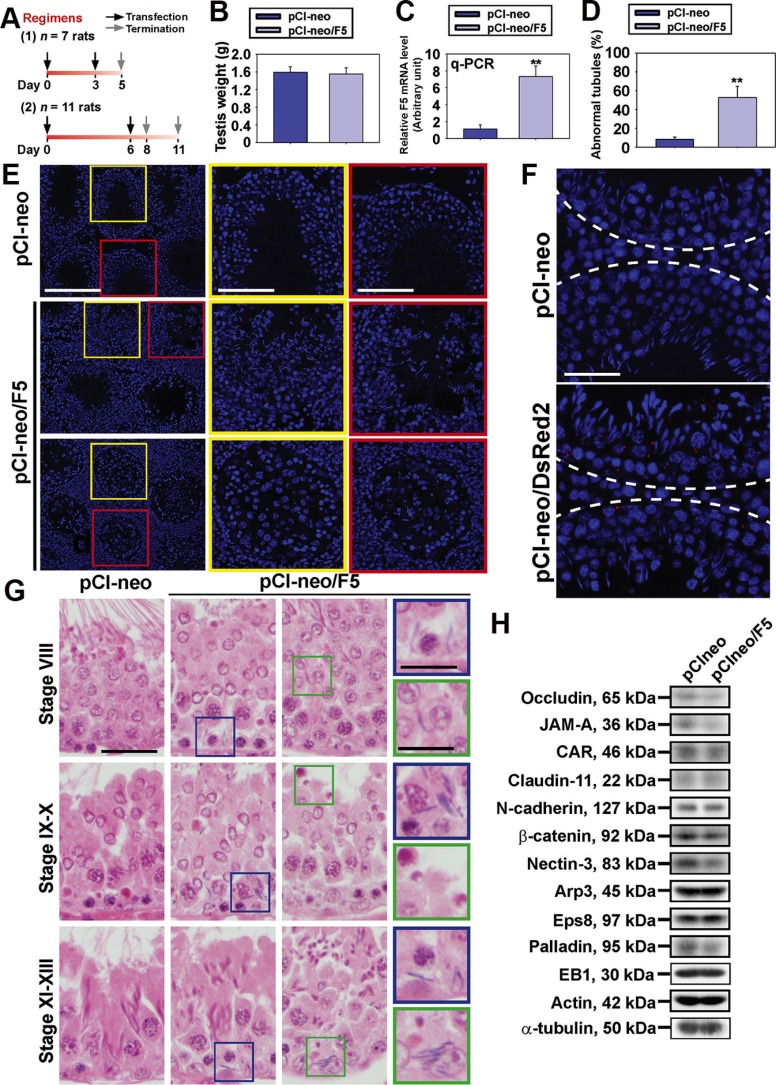
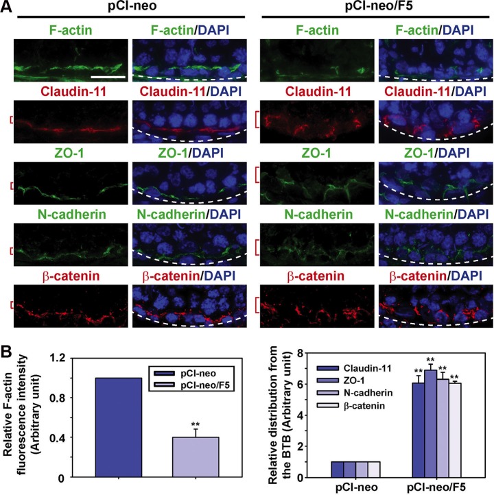
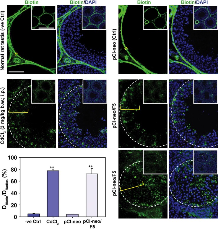
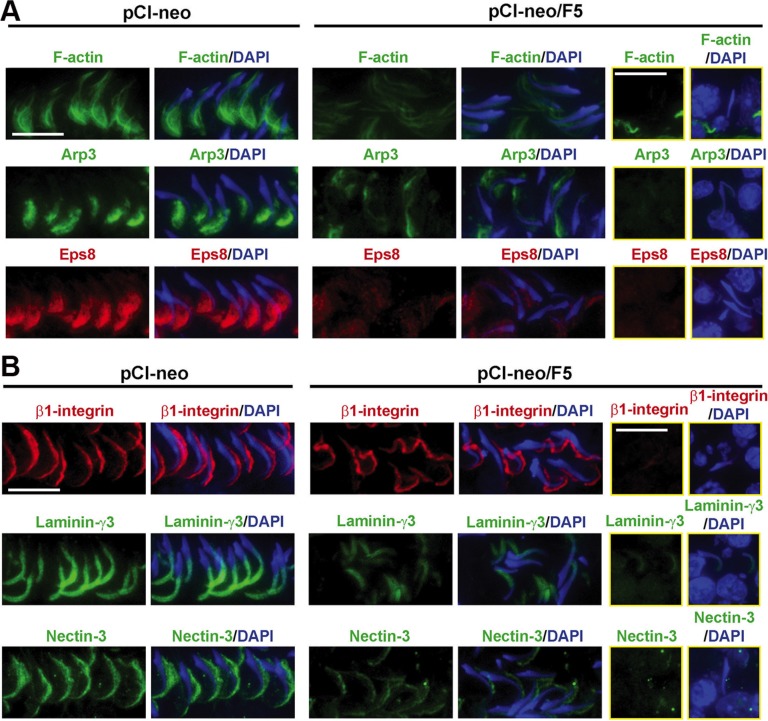
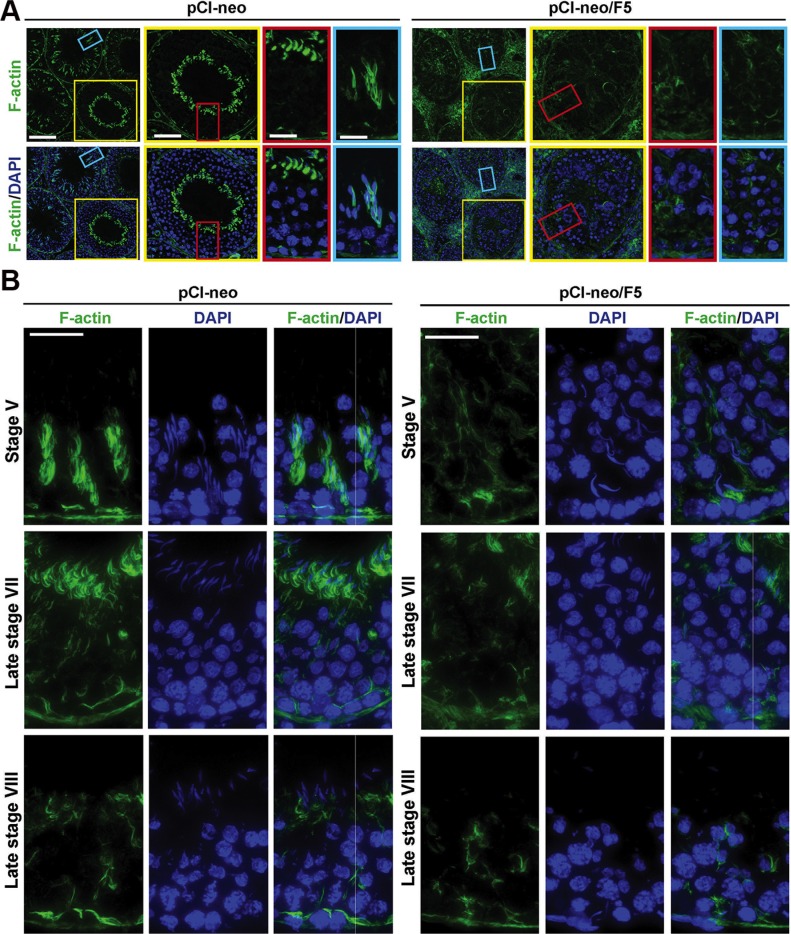
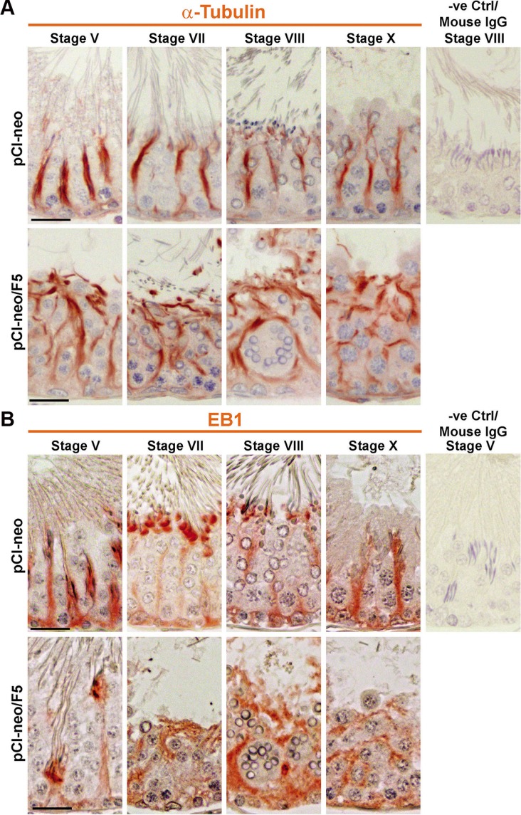
During the release of sperm at spermiation, a biologically active F5-peptide, which can disrupt the Sertoli cell tight junction (TJ) permeability barrier, is produced at the site of the degenerating apical ES (ectoplasmic specialization). This peptide coordinates the events of spermiation and blood-testis barrier (BTB) remodeling at stage VIII of the epithelial cycle, creating a local apical ES-BTB axis to coordinate cellular events across the epithelium. The mechanism(s) by which F5-peptide perturbs BTB restructuring, and its involvement in apical ES dynamics remain unknown. F5-peptide, besides perturbing BTB integrity, was shown to induce germ cell release from the epithelium following its efficient in vivo overexpression in the testis. Overexpression of F5-peptide caused disorganization of actin- and microtubule (MT)-based cytoskeletons, mediated by altering the spatiotemporal expression of actin binding/regulatory proteins in the seminiferous epithelium. F5-peptide perturbed the ability of actin microfilaments and/or MTs from converting between their bundled and unbundled/defragmented configuration, thereby perturbing adhesion between spermatids and Sertoli cells. Since apical ES and basal ES/BTB are interconnected through the underlying cytoskeletal networks, this thus provides an efficient and novel mechanism to coordinate different cellular events across the epithelium during spermatogenesis through changes in the organization of actin microfilaments and MTs. These findings also illustrate the potential of F5-peptide being a male contraceptive peptide for men.

Keywords: F5-peptide; actin cytoskeleton; ectoplasmic specialization; microtubule cytoskeleton; testis.


Conflict of interest statement

CONFLICTS OF INTEREST

Nothing to declare.

Figures

Figure 1
Figure 1. F5-peptide perturbs distribution of BTB-associated proteins at the Sertoli cell-cell interface
Sertoli cells were cultured alone for 3 day, and transfected with pCI-neo/F5 vs. pCI-neo vector alone (control) for 24 hr. Thereafter, cells were rinsed with F12/DMEM and terminated 24 hr later for RT-PCR and IF on day 5 vs. 48 hr later for IB on day 6. (A) Successful overexpression of F5-peptide was confirmed by RT-PCR and q-PCR by using a specific primer pair (Table S2) specific to F5-peptide. S16 served as a loading control for RT-PCR. GAPDH served as an internal control for q-PCR. (B) The steady-state levels of TJ proteins, basal ES proteins, actin regulatory proteins, MT regulatory protein, and signaling proteins found at the BTB were analyzed by IB. Overexpression of F5-peptide caused down-regulation of actin bundling proteins palladin and plastin 3 and also TJ proteins occludin and JAM-A. Actin served as a loading control. Histograms on the lower panel illustrate the down-regulation of occludin, JAM-A, palladin and plastin 3 following overexpression of F5-peptide. Each bar is a mean ± SD of n = 5 experiments, and data were normalized against actin. **P < 0.01. (C) Localization of TJ proteins (green fluorescence) CAR and ZO-1; and basal ES proteins N-cadherin and β-catenin; were analyzed by IF. Overexpression of F5-peptide caused a considerable reduction of CAR and ZO-1 fluorescence (see white (Ctrl, pCI-neo) vs. yellow (pCI-neo/F5) rectangle boxes) at cell-cell interface, whereas N-cadherin and β-catenin were shown to be diffusely localized at the cell cortical zone (see white (Ctrl, pCI-neo) vs. yellow (pCI-neo/F5) brackets) but internalized. Sertoli cell nuclei were visualized by DAPI (blue). The red fluorescence of Cy3-labeled plasmid DNA confirmed the successful transfection. Histograms on the lower panel summarize the findings on the upper panel to illustrate the loss of CAR/ZO-1 vs. re-distribution of N-cadherin/β-catenin at or from the cell-cell interface, respectively. Each bar is a mean ± SD of n = 3 experiments. **P < 0.01. Scale bar, 30 μm, which applies to all micrographs.
Figure 2
Figure 2. Overexpression of F5-peptide induces re-organization of actin- and microtubule (MT)-based cytoskeletons in Sertoli cells
(A) Overexpression of F5-peptide induced disorganization of actin microfilaments in which truncation of F-actin network (green or gray) in Sertoli cells was noted. Scale bar, 30 μm, which applies to other micrographs. (B) Actin bundling assay was performed to illustrate overexpression of F5-peptide led to a considerable loss of actin bundling capability. In this biochemical assay, pellet contained bundled actin microfilaments, whereas supernatant contained unbundled, linear and/or truncated actin microfilaments. Histograms summarize the findings on the upper panel, and each bar is a mean ± SD of n = 4 experiments. **P < 0.01. (C) Overexpression of F5-peptide caused internalization of branched actin polymerization protein Arp3. Moreover, actin barbed end capping and bundling protein Eps8 was diffusely localized at the cell-cell interface. These changes thus led to the generation of branched and truncated actin microfilaments in Sertoli cell cytosol as noted in (A). Scale bar, 30 μm. (D) F5-peptide also impaired microtubule (MT) organization when MT was visualized by α-tubulin staining which is the building block of MTs. Following overexpression of F5-peptide, α-tubulin-based MTs no longer stretched across the entire Sertoli cells as noted in control cells, instead, MTs were found to round up, encircling the Sertoli cell nuclei. Scale bar, 30 μm. Sertoli cell nuclei were visualized by DAPI (blue). In A, C and D, Cy3-labeled plasmid DNAs (red) illustrate successful transfection.
Figure 3
Figure 3. Overexpression of F5 peptide impairs spermatogenesis in vivo
(A) Two regimens were used since phenotypes on day 5 (Regimen 1) vs. day 8 and 11 (Regimen 2) were similar, data were pooled for analysis. (B) Testis weight in control (pCI-neo) vs. pCI-neo/F5 in both regimens of n = 18 rats. (C) F5-peptide mRNA level was analyzed by q-PCR. GAPDH served as an internal control. Each bar is a mean ± SD of n = 9 rats. **P < 0.01. (D) Percentage of damaged tubule was assessed using frozen sections of testes (see Materials and Methods). Each bar is a mean ± SD of n = 4 rats. **P < 0.01. (E) Morphological analysis using frozen sections stained with DAPI. Boxed areas in yellow or red were magnified and shown in insets. Germ cell loss was detected in tubules within 48 hr following the last transfection to overexpress F5-peptide. Scale bar in first column, 150 μm; 70 μm in yellow and red boxed micrographs. (F) Transfection efficiency was assessed by transfecting rat testes (n = 3 rats) with pCI-neo vs. pCI-neo/DsRed2 using Regimen 1. Positive transfection was confirmed by > 10 DsRed2 red fluorescence aggregates in the epithelium. Scale bar, 30 μm. (G) Histological analysis using paraffin sections of testes. In control testes transfected with pCI-neo empty vector, elongated spermatids line-up near the tubule lumen edge in stage VIII tubules to prepare for spermiation, but elongated spermatids remain entrapped inside the epithelium in stage VIII tubules after F5-peptide overexpression. At stages IX-X and XI-XIII tubules in control testes, no step 19 elongated spermatids were found in the epithelium, and phagosomes were transported to the base of epithelium. However, similar staged tubules following F5-peptide overexpression led to retention of elongated spermatids embedded deep inside the epithelium, and phagosomes were found near the luminal edge (see blue or green boxed area). Scale bar in the first column, 30 μm, which applies to 2nd/3rd column; scale bar in the last column, 10 μm. (H) Steady-state levels of the TJ-, basal/apical ES-, and actin or MT regulatory proteins were analyzed by IB with actin and α-tubulin served as loading controls. These are representative findings of n = 9 rats.
Figure 4
Figure 4. Overexpression of F5-peptide perturbs distribution TJ and basal ES proteins at the BTB through changes in the organization of F-actin in adult rat testes in vivo
(A) Frozen sections obtained from testes transfected with pCI-neo/F5 vs. pCI-neo alone (control) were stained for F-actin, TJ proteins claudin-11 and ZO-1, as well as basal ES proteins N-cadherin and β-catenin. Cell nuclei were visualized by DAPI (blue). The fluorescence intensity of F-actin at the BTB was considerably diminished after F5-peptide overexpression, consistent with in vitro observation due to truncation and dis-organization of actin microfilaments at the site. TJ proteins claudin-11 and ZO-1, as well as basal ES proteins N-cadherin and β-catenin were found to be diffusely localized at the Sertoli cell BTB in the testis transfected with pCI-neo/F5, supporting findings in vitro that illustrate an increase in protein internalization at the site. Basement membrane was annotated by a dashed white line illustrating the relative location of the basement membrane/tunica propria. Scale bar, 30 μm, which applies to all other micrographs. (B) Semi-quantitative analysis of fluorescence data shown in (A) including fluorescence intensity (left panel) vs. mis-localization of TJ and basal ES proteins by diffusing away from the BTB (right panel). Data in the control group were arbitrarily set at 1 against which statistical comparison was performed. Each bar is a mean ± SD of n = 4 rats. **P < 0.01.
Figure 5
Figure 5. Overexpression of F5-peptide in the test is perturbs BTB function in vivo
BTB integrity assay was performed to assess the ability of an intact BTB to block the diffusion of a small molecule biotin (EZ-Link Sulfo-NHS-LC-Biotin, Mr 556.59) across the BTB which biotinylated proteins in the adluminal compartment. In normal rat testes and testes transfected with pCI-neo alone (control), the functional BTB blocked biotin reagent from entering the adluminal compartment so that only proteins at the BTB and the interstitial space were labeled. Biotinylated proteins were subsequently visualized using frozen sections of the testis and stained with Alexa Fluor 488-streptavidin (green fluorescence). Distance traveled by biotin beyond the basement membrane/tunica propria was annotated by a yellow bracket. In control testes, biotin failed to travel beyond the basement membrane/tunica propria which was annotated by a dashed white line. In testes from rats treated with CdCl2 (positive control) or transfected with pCI-neo/F5, biotin penetrated well into the adluminal compartment, illustrating that F5-peptide perturbed the BTB integrity. Lower magnified images of ~3–4 tubules were shown in insets and the enlarged image of a typical tubule was shown in the micrograph. Scale bars, 50 μm, and 200 μm in insets, which applies to corresponding micrographs and/or insets. Histograms show the semi-quantitative data by comparing the distance of biotin traveled into the epithelium (DBiotin) vs. the radius of seminiferous tubule (DRadius). For oblique sections, the radius of the tubule was obtained by averaging the longest and shortest distance from the basement membrane. Each bar is a mean ± SD of ~50 tubules that were randomly selected and scored from testes of 6 rats with a total of 300 randomly selected tubules. **P < 0.01.
Figure 6
Figure 6. Overexpression of F5-peptide perturbs F-actin organization through changes in the spatial expression of actin regulatory proteins, which in turn disrupts apical ES protein distribution in adult rat testes in vivo
Frozen sections of testes following transfection of pCI-neo/F5 vs. pCI-neo (vector alone, control) were stained for F-actin, Arp3, and Eps8; as well as integral membrane proteins at the apical ES: β1-integrin (Sertoli cell-specific) vs. spermatid-specific laminin-γ3 chain and nectin-3. Spermatids that were entrapped inside the seminiferous epithelium were shown in the two rectangular columns on the right. Cell nuclei were visualized by DAPI. (A) At stage VII, F-actin, actin regulatory proteins Arp3 and Eps8 were prominently localized to the concave side of spermatid heads in control testes. Overexpression of F5-peptide, however, induced mis-organization of F-actin which was considerably diminished at the apical ES found in elongated spermatids located near the tubule lumen or embedded inside the epithelium. These changes in F-actin organization were the result of changes in spatial expression of Arp3 and Eps8 since these proteins no longer restricted to the concave side of spermatids, and their expression was considerably diminished. Furthermore, many spermatids had lost their polarity since they no longer pointed toward the basement membrane as found in control tubules. For spermatids entrapped inside the epithelium in these tubules, the fluorescence signals of Arp3 and Eps8 were also considerably diminished after F5-peptide overexpression, illustrating apical ES in these spermatids was also disrupted. Scale bar, 10 μm, which applies to other micrographs. (B) Apical ES adhesion proteins β1-integrin and nectin-3 were localized at the convex side of spermatid heads whereas laminin-γ3 chain was at the tip of spermatid heads in normal testes. Following F5-peptide overexpression, β1-integrin was grossly mis-localized since some fluorescence signal was found on the concave side of spermatid heads; whereas laminin-γ3 chain and nectin-3 were also considerably down-regulated and mis-localized, thereby impeding spermatid adhesion, leading to germ cell exfoliation as noted in Figure 3. Scale bar, 10 μm, which applies to other micrographs in the same panel.
Figure 7
Figure 7. Overexpression of F5-peptide perturbs F-actin organization in the seminiferous epithelium of adult rat testes in vivo
Rat testes were transfected with pCI-neo/F5 vs. pCI-neo (control) using Regimens (n = 9 rats) shown in Figure 3A. Frozen sections of testes were used to visualize the distribution of F-actin by FITC-phalloidin. Cell nuclei were visualized by DAPI. (A) Overexpression of F5-peptide led to extensive germ cell loss in tubules wherein F-actin distribution was grossly disrupted. The typical “stalk-like” structures of F-actin was not found in tubules following overexpression with F5-peptide. Scale bar in the left column, 100 μm; Scale bar in yellow box, 50 μm; red or blue box, 15 μm. (B) Representative micrographs of frozen sections of normal testes in selected stages were stained for F-actin. As noted herein, F-actin appeared as “stalk-like” structures, tightly associated with elongating spermatids in transport across the epithelium in stage V tubules, also associated with apical ES surrounding the head of elongated spermatids in late stage VII, but considerably diminished in late stage VIII tubules to facilitate the release of sperms at spermiation. Moreover, some stalk-like structure reappeared across the epithelium in late stage VIII, perhaps being used to support the transport of residual bodies/phagosome to the basal compartment for their eventual lysosomal degradation. However, following F5-peptide overexpression, the typical F-actin organization in the epithelium was considerably disrupted, For instance, elongating spermatids in stage V tubules no longer associated with the F-actin-based stalk-like structures and F-actin was considerably diminished in stage VII-VIII tubules. Elongated spermatids, however, remained entrapped inside the epithelium even though the apical ES had been disrupted. Scale bar, 25 μm, which applies to other micrographs.
Figure 8
Figure 8. Overexpression of F5-peptide perturbs MT organization in the seminiferous epithelium of adult rat testes in vivo
Rat testes were transfected with pCI-neo/F5 vs. pCI-neo empty vector (control) using regimens (n = 9 rats) shown in Figure 3A. Testes were used for immunohistochemistry to visualize the distribution of α-tubulin, the building block of microtubules (MTs) in the seminiferous epithelium. (A) In normal testes, MTs appeared as track-like structures were found longitudinally across the epithelium to provide the tracks to support the transport of organelles (e.g., phagosomes, endocytic vesicles) and also spermatids in virtually all stages of the epithelial cycle. Following overexpression of F5-peptide, on day 2 or day 5 after completion of transfection, tracks were grossly affected. They no longer distinctively found across the epithelium, but truncated and some laid across the epithelium in parallel to the tunica propria. Moreover, MTs were also found to engulf round spermatids, becoming the precursors of giant multinucleated round spermatids, destined to be degenerated. The last micrograph on the right of the top panel is the negative control (a stage VIII tubule) in which the primary antibody was substituted with the mouse IgG. Scale bar, 30 μm, which applies to other micrographs. (B) Changes in the organization of EB1, a +TIP protein known to bind to MTs, helping to stabilize MTs, were also noted in testes following overexpression of F5-peptide. Similar to α-tubulin, EB1 appeared as track-like structures across the epithelium, but overexpression of F5-peptide led to either considerably loss of EB1 in the epithelium, and it no longer laid across the epithelium longitudinally, but truncated and also wrapped around groups of round spermatids destined to become giant multinucleated round spermatids for eventual degeneration. The last micrograph on the right of the top panel is the negative control (a stage V tubule) in which the primary antibody was substituted with the mouse IgG. Scale bar, 30 μm, which applies to other micrographs.

Similar articles

Cited by

References

    1. Cheng CY, Mruk DD. A local autocrine axis in the testes that regulates spermatogenesis. Nature Rev Endocrinol. 2010;6:380–395. - PMC - PubMed
    1. Cheng CY, Mruk DD. An intracellular trafficking pathway in the seminiferous epithelium regulating spermatogenesis: a biochemical and molecular perspective. Crit Rev Biochem Mol Biol. 2009;44:245–263. - PMC - PubMed
    1. Yan HHN, Mruk DD, Wong EWP, Lee WM, Cheng CY. An autocrine axis in the testis that coordinates spermiation and blood-testis barrier restructuring during spermatogenesis. Proc Natl Acad Sci USA. 2008;105:8950–8955. - PMC - PubMed
    1. Wong EWP, Cheng CY. NC1 domain of collagen α3(IV) derived from the basement membrane regulates Sertoli cell blood-testis barrier dynamics. Spermatogenesis. 2013;3:e25465. - PMC - PubMed
    1. Su L, Mruk DD, Lie PPY, Silvestrini B, Cheng CY. A peptide derived from laminin-γ3 reversibly impairs spermatogenesis in rats. Nat Communs. 2012;3:1185. - PMC - PubMed

MeSH terms

LinkOut - more resources